- Veterinary View Box
- Posts
- 3T MRI Flag for Headshaking: Trigeminal Asymmetry Pinpoints ITMHS
3T MRI Flag for Headshaking: Trigeminal Asymmetry Pinpoints ITMHS
JVIM 2025
Frederik Heun, Julien Delarocque, Karsten Feige, Maren Hellige
Background
Idiopathic trigeminal-mediated headshaking (ITMHS) is a prevalent and painful equine disorder whose pathophysiology remains uncertain. Human trigeminal neuralgia literature suggests that unilateral trigeminal nerve (TN) changes—including atrophy—may be detectable on MRI; however, comparable MRI-based morphometry had not been evaluated in horses. This study tested whether horses with ITMHS exhibit greater side-to-side asymmetry of TN cross-sectional area (TNCSA) than controls.
Methods
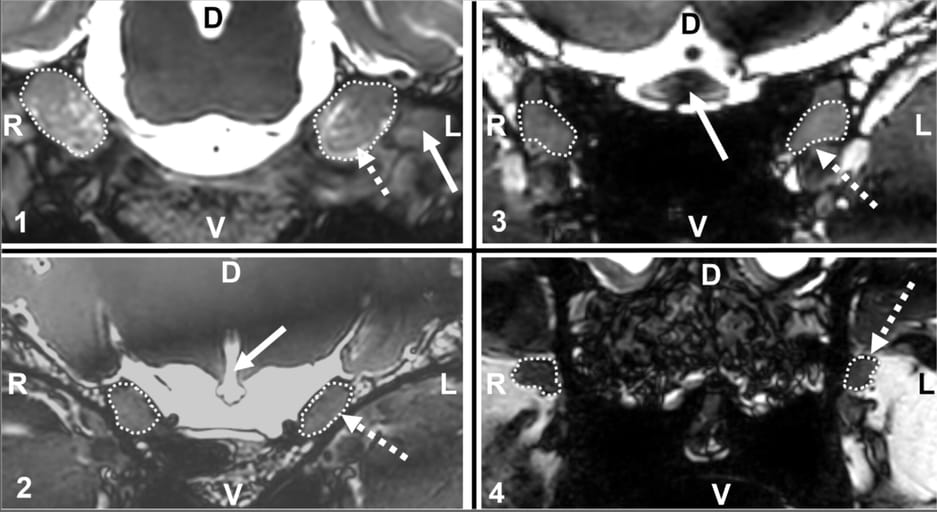
This retrospective case–control study analyzed 3-Tesla brain MRI from 20 adult horses with ITMHS and six controls scanned in 2021–2022 at a single equine referral center. TNCSA was measured bilaterally at four predefined measurement points (MP1–MP4) along the ophthalmic/maxillary course using standardized plane alignment and primarily 3D balanced gradient-echo sequences. A linear mixed model assessed effects of group and location on absolute TNCSA and on side-to-side TNCSA differences (primary outcome). Repeatability was tested by intraclass correlation coefficients (ICC). Potential effects of age and body weight were explored; data-driven cut-offs for TNCSA asymmetry at each MP were derived.
Results
Horses with ITMHS had significantly greater TNCSA asymmetry than controls (main effect p < 0.001), with 4.1–7.6-fold larger side-to-side differences across most locations (notably MP1, MP2, and MP4). Absolute TNCSA did not differ between groups but increased with body weight and varied by location (largest at MP1, smallest at MP4). Measurement repeatability was excellent (overall ICC ≈ 0.98). Suggested asymmetry cut-offs discriminated ITMHS from controls best at MP2 (Youden-optimized threshold ~1.45 mm²; perfect separation in this dataset), while MP3 performed poorly due to artifacts/variability. Duration of clinical signs did not correlate with asymmetry.
Limitations
The sample was modest with only six controls, and controls had intracranial disorders unlikely—but not guaranteed—not to affect TN morphology. All measurements were performed by a single, non-blinded examiner. MRI protocols, though standardized, faced susceptibility/band artifacts at MP3. The study lacked histopathology, and cut-offs were derived and tested on the same cohort, likely overestimating diagnostic performance.
Conclusions
MRI-detected TNCSA asymmetry is increased in horses with ITMHS, supporting unilateral or asymmetric TN involvement and highlighting MRI’s value in assessment. While absolute nerve size is confounded by body size, side-to-side asymmetry—particularly at MP2—emerges as a promising imaging biomarker. External validation and histologic correlation are needed, but the findings may inform future targeted (potentially unilateral) therapies.

Four images from a balanced gradient echo sequence of the brain of one horse with headshaking. The figure is subdivided into four quadrants: One for each measurement point (1–4). Each quadrant shows a transverse MRI image displaying the trigeminal nerves and surrounding tissue. The dotted circle indicates the trigeminal nerve cross-sectional area (measurement point). The left trigeminal nerve is indicated by the dotted arrow. Bold arrows indicate anatomical landmarks: (1) mandibular nerve, (2) infundibular recess, (3) optic chiasm, D, dorsal; L, left; R, right; V, ventral. Note the slight difference in cross-sectional area, for example at measurement point 2: right 50 mm2, left 61 mm2.
How did we do? |
Disclaimer: The summary generated in this email was created by an AI large language model. Therefore errors may occur. Reading the article is the best way to understand the scholarly work. The figure presented here remains the property of the publisher or author and subject to the applicable copyright agreement. It is reproduced here as an educational work. If you have any questions or concerns about the work presented here, reply to this email.