- Veterinary View Box
- Posts
- A basset disease or normal variant?
A basset disease or normal variant?
Journal of Small Animal Practice 2018
A. Lorek, S. E. Spencer, R. Dennis
Background
Masticatory myositis and other inflammatory myopathies can be diagnosed using magnetic resonance imaging (MRI), but normal breed variations in muscle appearance may lead to misinterpretation. Basset Hounds often exhibit characteristic MRI changes in masticatory muscles, which may be mistaken for disease. This study aimed to describe MRI characteristics of masticatory muscles in Basset Hounds to differentiate normal breed variations from pathological myopathies.
Methods
Study Design:
-Retrospective review of 44 Basset Hound MRI scans (high-field: 33 dogs; low-field: 11 dogs).
-Dogs were imaged for unrelated reasons (e.g., seizures, neurological deficits, otitis, exophthalmos).
Imaging Protocols:
-T1-weighted (T1W), T2-weighted (T2W), FLAIR, GRE, and STIR sequences.
-Pre- and post-contrast T1W imaging with and without fat suppression.
Analysis:
-Subjective grading of muscle bulk, signal intensity, and symmetry.
-Ordinal logistic regression assessed associations between age, weight, and MRI changes.
Results
-Muscle Changes Observed in 39/44 Dogs (88.6%)
-Reduced muscle bulk in the superficial temporalis and masseter muscles.
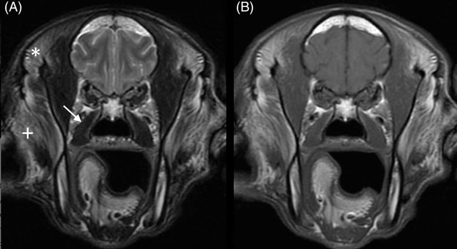
-Bilateral, symmetrical T1W and T2W hyperintensity, suggestive of fat infiltration.
-No contrast enhancement, confirming absence of inflammation.
Fat Suppression Findings:
-Signal reduction with STIR and post-contrast fat suppression, confirming fat replacement, not pathology.
-Medial pterygoid and digastricus muscles were unaffected, distinguishing this from masticatory myositis.
Age and Weight Correlation:
-Severity increased weakly with age and weight (p < 0.0001).
-Changes were stable over time, with no progression in follow-up MRIs.
Limitations
No muscle biopsies performed in most dogs (one biopsy confirmed fatty infiltration without inflammation).
Retrospective study design, leading to variations in imaging protocols.
No functional assessment (e.g., electromyography) to confirm nerve involvement.
Conclusions
The MRI characteristics of masticatory muscles in Basset Hounds are breed-specific variations, not pathological changes. The observed fatty infiltration and muscle bulk reduction are likely related to chronic mechanical strain from their heavy ears rather than inflammatory myopathy. Awareness of these breed-specific findings can prevent misdiagnosis and unnecessary invasive diagnostics in Basset Hounds.

Transverse T2W (A) and T1W (B) high-field image of the head of a 10-year-old basset hound with severe, bilaterally symmetrical hyperintensity in the superficial part of the temporalis muscles (*) and the dorsal aspect of the masseter muscles (+) as well as mild atrophy of the temporalis muscles. The parts of the temporalis muscles deep to the mandibular coronoid processes are spared. The pterygoid muscles appear normal (arrow)
How did we do? |
Disclaimer: The summary generated in this email was created by an AI large language model. Therefore errors may occur. Reading the article is the best way to understand the scholarly work. The figure presented here remains the property of the publisher or author and subject to the applicable copyright agreement. It is reproduced here as an educational work. If you have any questions or concerns about the work presented here, reply to this email.