- Veterinary View Box
- Posts
- Advancing Equine Tendon Imaging: CT Tenography of the Carpal Flexor Sheath
Advancing Equine Tendon Imaging: CT Tenography of the Carpal Flexor Sheath
Veterinary Radiology & Ultrasound 2025
Thomas David Chisholm Woods, Jonathon Dixon, Barny Simon Lovat Fraser, Chris Melvaine
Background
The equine carpal flexor tendon sheath (CFTS) envelops critical tendinous and neurovascular structures in the palmar carpal region. Pathologies affecting the CFTS, such as tenosynovitis or flexor tendon injuries, are challenging to diagnose due to anatomical complexity. Exploratory tenoscopy is the current gold standard for evaluating intrathecal lesions; however, preoperative imaging could improve prognostication and surgical planning. This study aimed to describe the normal anatomy of the equine CFTS using noncontrast and contrast-enhanced computed tomographic (CT) tenography in cadaver limbs.
Methods
Ten pairs of cadaveric equine forelimbs from clinically normal adult horses were examined using an 80-slice multidetector CT scanner. Each specimen underwent a noncontrast CT scan followed by an injection of iohexol contrast medium into the CFTS, with subsequent contrast-enhanced CT imaging. Multi-planar reformatting was used to assess the visibility of intrathecal and extrathecal structures. Tenoscopy and gross dissection were performed to confirm findings and identify anatomical variations.
Results
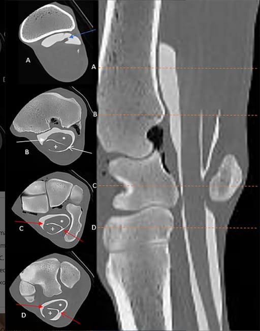
Noncontrast CT successfully identified major structures, including the superficial and deep digital flexor tendons (SDFT, DDFT), accessory ligaments, and suspensory ligament, with contrast-enhanced CT improving visualization of finer details. Newly documented structures, such as the proximal mesotenon and the manica of the common mesotenon, were identified. The distal termination of the CFTS was found to extend further distally than previously reported, often beyond the mid-metacarpus, highlighting potential risks for iatrogenic sheath penetration during diagnostic anesthesia or surgical procedures. No significant pathology was detected in the specimens, though anatomical variations such as an intrathecal ulnar head of the DDFT were noted in some limbs.
Limitations
The study used a small sample of Standardbred horses, limiting the generalizability of findings to other breeds. Postmortem changes may have affected soft tissue integrity, and the study did not assess clinical pathology cases. Further research is needed to evaluate the sensitivity and specificity of CT tenography in diagnosing tendon sheath disease in live horses.
Conclusions
CT tenography provides detailed visualization of the equine CFTS, surpassing conventional imaging modalities in anatomical clarity. The identification of previously undescribed mesotenons enhances the understanding of tendon sheath anatomy and may influence future surgical and diagnostic approaches. The extended distal termination of the CFTS raises considerations for anesthesia administration and surgical access. These findings support further clinical application of CT tenography for preoperative planning and improved diagnostic accuracy in equine musculoskeletal medicine.

Transverse and sagittal contrast CT images in bone window showing the radial head of the deep digital flexor tendon (A; blue arrow), the proximal mesotenon (B; white arrows, shaded arrowhead is medial) and the manica of the common mesotenon (C, D; red arrows, shaded arrowhead is medial, site of attachment to the common mesotenon). N.B. The dashed orange lines correspond to the transverse sections on the left (images A–D); * is the deep digital flexor tendon, + is the superficial digital flexor tendon.
How did we do? |
Disclaimer: The summary generated in this email was created by an AI large language model. Therefore errors may occur. Reading the article is the best way to understand the scholarly work. The figure presented here remains the property of the publisher or author and subject to the applicable copyright agreement. It is reproduced here as an educational work. If you have any questions or concerns about the work presented here, reply to this email.