- Veterinary View Box
- Posts
- AI can help....
AI can help....
Veterinary Radiology & Ultrasound 2025
Mai, W.; Hecht, S.; Paek, M.; Holmes, S.P.; Dorez, H.; Blanchard, M.; Nour Eddin, J.
Background:
Magnetic resonance imaging (MRI) in veterinary medicine is limited by long scan times and inherent noise, which can reduce image quality. Strategies such as high-field MRI scanners and advanced reconstruction techniques exist but are expensive or scanner-specific. Deep learning-based denoising algorithms have been developed for human imaging, but a validated, veterinary-specific solution is lacking. This study evaluated the effectiveness of a novel DICOM-based deep learning (DL) denoising algorithm for improving MRI image quality in veterinary patients.
Methods:
Thirty canine and feline brain MRI scans were analyzed, comparing native images with those processed using the HawkAI DL denoising algorithm. Signal-to-noise ratio (SNR) and contrast-to-noise ratio (CNR) were quantitatively measured on T2-weighted (T2W), T2-FLAIR, and Gradient Echo (GRE) sequences. Three board-certified veterinary radiologists conducted blinded qualitative assessments of image coarseness, contrast, and overall quality. Statistical analysis included Wilcoxon signed-rank tests to compare native and AI-processed images.
Results:
SNR and CNR were significantly higher in DL-processed images compared to native images across all MRI sequences (p < 0.0001). The improvement in SNR ranged from 88.1% to 94.4% for T2W, 34.9% to 40.7% for T2-FLAIR, and 38.1% to 41.8% for GRE images. CNR improved by 99.7% to 105.2% for T2W, 44.4% to 48.6% for T2-FLAIR, and 37.7% to 45.7% for GRE images. Subjective image quality scores also improved significantly, particularly for T2W and GRE sequences, with better perceived contrast and reduced coarseness.
Limitations:
The study was limited by a small sample size and the use of a single MRI scanner model, requiring further validation across different imaging platforms. The algorithm was only assessed for noise reduction and not its clinical impact on diagnosis. The study design did not include side-by-side qualitative comparisons, which might have provided stronger subjective assessments.
Conclusions:
The veterinary-specific DL-based denoising algorithm significantly improved both objective and subjective image quality in 1.5T MRI scans of dogs and cats. These findings suggest its potential for reducing scan times or enhancing diagnostic confidence in veterinary MRI. Future research should explore its applicability across various MRI platforms and its clinical impact on diagnostic accuracy.

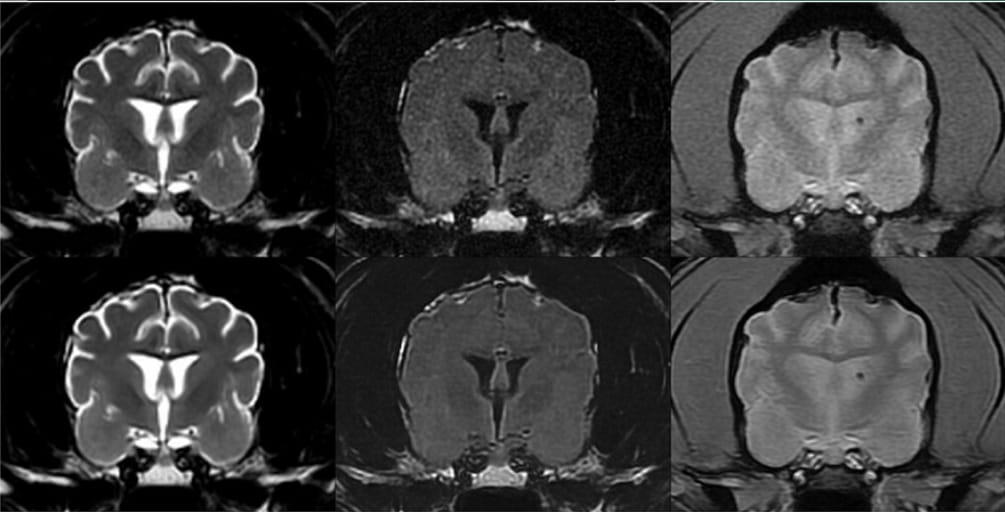
Transverse T2W (left), T2-FLAIR (middle), and GRE (right) brain MRI images in a 13-year-old standard poodle. Native images are at the top, and corresponding AI-treated images are at the bottom. For this patient, the increase in SNR on AI-treated images ranged between 77% and 109% for the T2W images, 24.6% and 37.3% for the T2-FLAIR images, and 61.9% and 84.7% for the GRE images. This patient received higher (i.e., better) coarseness, contrast, and overall quality scores for the AI-treated images than the native images. A left-sided microbleed is visible on the GRE images. Slice thickness = 3 mm, interslice gap = 0.5 mm, FOV = 14 cm; T2W image parameters: TR = 3665 ms, TE = 100 ms, NEX = 3; T2-FLAIR image parameters: TR = 8962 ms, TE = 146 ms, TI = 2376 ms, NEX = 2; GRE image parameters: TR = 717 ms, TE = 15 ms, Flip angle = 20°, NEX = 2.
How did we do? |
Disclaimer: The summary generated in this email was created by an AI large language model. Therefore errors may occur. Reading the article is the best way to understand the scholarly work. The figure presented here remains the property of the publisher or author and subject to the applicable copyright agreement. It is reproduced here as an educational work. If you have any questions or concerns about the work presented here, reply to this email.