- Veterinary View Box
- Posts
- Anatomy of the right outflow tract....
Anatomy of the right outflow tract....
Veterinary Radiology & Ultrasound 2025
Chiara Massarenti, Marta Croce, Alessia Diana, Massimiliano Tursi, Eric Zini, Oriol Domenech, Edoardo Auriemma
Background
Evaluation of the pulmonary valve and right ventricular outflow tract (RVOT) in dogs relies primarily on echocardiography, but its two-dimensional nature and limitations in certain breeds (e.g., brachycephalic dogs) can hinder diagnostic accuracy. In human medicine, ECG-gated multidetector computed tomography (MDCT) provides detailed cardiac imaging. This study aimed to describe ECG-gated MDCT findings of the normal pulmonary valve and RVOT in dogs to establish baseline anatomical reference values.
Methods
Study Design:
Prospective anatomical descriptive study conducted at the AniCura Istituto Veterinario Novara and the University of Turin.
Study Population:
-24 client-owned dogs without pulmonary valve or RVOT abnormalities.
-Three additional cadaveric canine hearts were used for gross and histological comparisons.
Imaging Protocol:
-All dogs underwent echocardiography before inclusion to exclude cardiac abnormalities.
-64-MDCT scanner (ECG-gated cardiac imaging) was used.
-Multiplanar reconstructions assessed four key anatomical structures:
-Pulmonary valve leaflets
-Sinotubular junction
-Anatomic ventriculo-arterial junction
-Hemodynamic ventriculo-arterial junction
Qualitative Assessment:
-Image quality was graded from 0 (not visible) to 2 (excellent visibility).
-Two independent observers (radiology resident and board-certified radiologist) reviewed the images.
Results
Visualization Quality of Pulmonary Valve Anatomy:
-Pulmonary valve leaflets were clearly visible in 88% of cases.
-Sinotubular junction (92% good visibility) and hemodynamic junction (88%) were well-defined.
-Anatomic ventriculo-arterial junction had the lowest visibility (75%), likely due to its complex structure.
Pulmonary Valve Morphology:
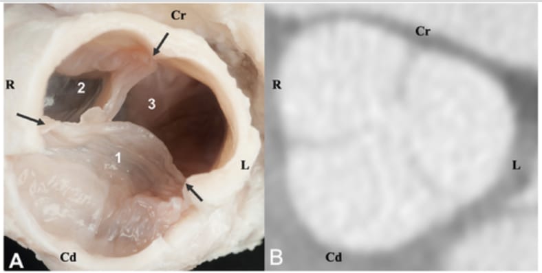
-The short-axis view of the pulmonary valve consistently showed a “Mercedes-Benz sign”, similar to the aortic valve appearance in diastole.
Comparison with Cadaveric Hearts:
-The CT images closely matched gross and histologic findings, confirming anatomical accuracy.
Limitations
Small sample size (n = 24), limiting generalizability.
No evaluation of diseased pulmonary valves, restricting clinical applicability.
Some cases had minor motion artifacts due to arrhythmias, affecting image clarity.
Conclusions
This study provides the first detailed CT description of the normal pulmonary valve and RVOT anatomy in dogs, demonstrating that ECG-gated MDCT offers excellent visualization of key cardiac structures. The "Mercedes-Benz sign" on CT may serve as a reference for normal pulmonary valve morphology. These findings support the potential clinical application of MDCT for diagnosing pulmonary valve disorders in veterinary medicine.

A, Photograph of the gross anatomy of a dorsal view of the pulmonary valve after removal of the lateral wall of the infundibulus in a 10-year-old Springer Spaniel (case no. 23) illustrating the left pulmonary cusp (1), the right pulmonary cusp (2), and intermediate pulmonary cusp (3). Black arrows indicate sinotubular junction. B, Dorsal multiplanar reformatted image showing the pulmonary valve at the same level of the anatomic image. R: right. L: left. Cr: cranial. Cd: caudal
How did we do? |
Disclaimer: The summary generated in this email was created by an AI large language model. Therefore errors may occur. Reading the article is the best way to understand the scholarly work. The figure presented here remains the property of the publisher or author and subject to the applicable copyright agreement. It is reproduced here as an educational work. If you have any questions or concerns about the work presented here, reply to this email.