- Veterinary View Box
- Posts
- Another staffy disease.....
Another staffy disease.....
JSAP 2015
Magnetic resonance imaging of suspected idiopathic bilateral C2 hypertrophic ganglioneuritis in dogs
S. Joslyn, C. Driver, F. McConnell, J. Penderis, A. Wessmann
Background
Hypertrophic ganglioneuritis is an inflammatory condition affecting nerve roots, commonly described in human medicine but rarely reported in dogs. While cervical nerve root enlargement is often attributed to tumors or infectious neuritis, cases of idiopathic hypertrophic ganglioneuritis remain poorly characterized. This study aimed to describe the MRI and clinical features of suspected idiopathic bilateral C2 hypertrophic ganglioneuritis in dogs, focusing on imaging characteristics, clinical presentation, and response to treatment.
Methods
Study Design: Retrospective review of MRI records from four veterinary referral institutions (2009–2012).
Inclusion Criteria:
-Dogs with bilateral C2 nerve root enlargement on MRI.
-Available clinical history, neurological assessment, and laboratory results.
Imaging Protocol:
-MRI scans (1T or 1.5T units) included sagittal, transverse, and dorsal T1-weighted (T1W) and T2-weighted (T2W) sequences.
-Post-contrast gadolinium-enhanced imaging was used to assess nerve root enhancement.
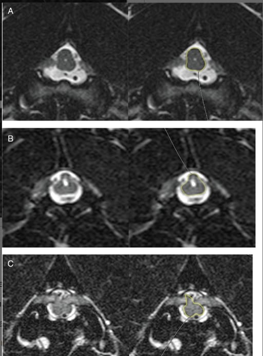
-Spinal cord compression was graded as mild, moderate, or severe based on shape distortion.
Results
Study Population:
-12 dogs met inclusion criteria.
-Nine were Staffordshire Bull Terriers, indicating a possible breed predisposition.
Clinical Signs and Neurological Findings:
-Eight dogs had clinically significant lesions (moderate-to-severe spinal cord compression).
-The remaining four dogs had incidental findings and presented for other neurological conditions (e.g., seizures, intervertebral disc disease).
-Most affected dogs exhibited cervical pain, ataxia, and proprioceptive deficits.
MRI Findings:
-Bilateral contrast-enhancing C2 nerve root enlargement was observed in all cases.
-Spinal cord compression ranged from mild to severe and correlated with neurological deficits.
-T2W hyperintensity was seen in four cases, possibly indicating edema or inflammation.
-No evidence of neoplasia or infectious causes was identified.
Treatment and Outcomes:
-Five of the eight clinically affected dogs received corticosteroids (immunosuppressive doses of prednisolone). All showed clinical improvement.
-Two untreated dogs improved with rest alone.
-One dog remained symptomatic, likely due to concurrent spinal disease.
Limitations
Retrospective study design, limiting control over MRI protocols and treatment regimens.
No histopathologic confirmation, preventing definitive classification of the condition as ganglioneuritis.
Small sample size, making statistical associations difficult.
Conclusions
Idiopathic bilateral C2 hypertrophic ganglioneuritis appears to be a distinct inflammatory neuropathy in dogs, with Staffordshire Bull Terriers overrepresented. MRI findings of bilateral, contrast-enhancing C2 nerve root enlargement should raise suspicion for this condition, particularly in dogs presenting with cervical myelopathy. Immunosuppressive corticosteroid therapy led to clinical improvement, suggesting a potential inflammatory or immune-mediated etiology. Further studies, including histopathology and genetic investigations, are needed to clarify the pathogenesis and optimal treatment strategies for this condition.

(A) Transverse T2W image at the level of the C1-2 intervertebral foramen. Mild distortion is present light lateral shape change to the spinal cord, highlighted with a region of interest. (B) Transverse T2W image at the level of the C1-2 intervertebral foramen. Moderate spinal cord ccausing triangular distortion of the spinal cord highlighted by the region of interest. The enlarged nerve roots are less than half the diameter of the cranial cervical spinal cord. (C) Transverse T2W image at the level of the C1-2 intervertebral foramen. Severe spinal cord compression and irregular shape change highlighted by the region of interest. The C2 nerve roots are thicker than half the diameter of the normal cranial cervical spinal cord
How did we do? |
Disclaimer: The summary generated in this email was created by an AI large language model. Therefore errors may occur. Reading the article is the best way to understand the scholarly work. The figure presented here remains the property of the publisher or author and subject to the applicable copyright agreement. It is reproduced here as an educational work. If you have any questions or concerns about the work presented here, reply to this email.