- Veterinary View Box
- Posts
- Are Your Swallow Studies Safe? New Data on Radiation Exposure in Veterinary Fluoroscopy
Are Your Swallow Studies Safe? New Data on Radiation Exposure in Veterinary Fluoroscopy
VRU 2025
Kathleen M. Lehman, Savannah R. Saboda, Kim R. Love, Kursten V. Pierce
Background
Videofluoroscopic swallow studies (VFSS) are the gold standard for diagnosing dysphagia in dogs and cats, yet they expose veterinary personnel to ionizing radiation. Despite the widespread use of VFSS in teaching hospitals and some private practices, data on occupational exposure during these procedures in veterinary medicine are lacking. This study aimed to document the radiation dose received by personnel during contrast-enhanced VFSS, evaluate exposure differences by disease localization, and assess the effect of operator training level on dose.
Methods
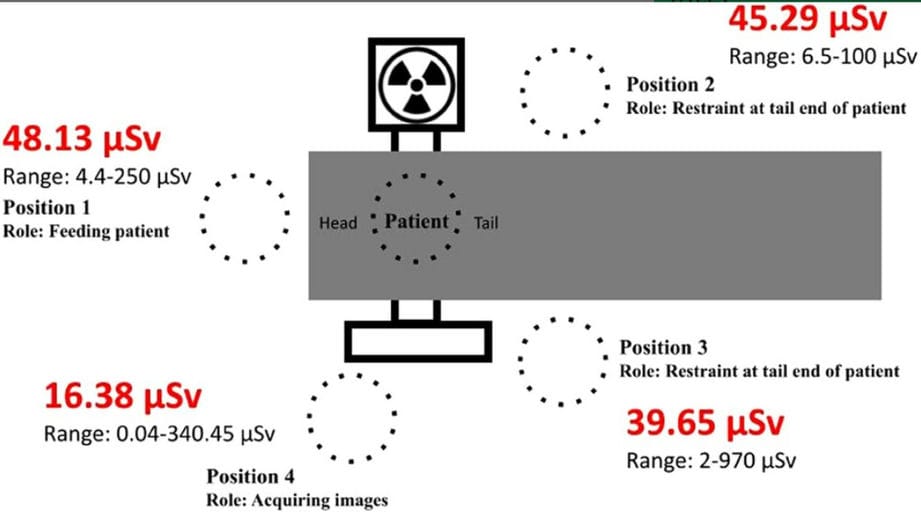
A prospective observational study was conducted at North Carolina State University’s Veterinary Teaching Hospital from October 2022 to May 2023. Sixty-one client-owned animals (58 dogs, 3 cats) underwent VFSS. Personnel wore dosimeters at four standardized positions:
-Position 1: Feeding/at patient’s head
-Positions 2 & 3: Restraining at tail end
-Position 4: Acquiring images
Fluoroscopy parameters, patient data, and procedure duration were recorded. Radiation exposure was analyzed using linear mixed-effects models, considering fluoroscopy time, patient weight, disease localization, and personnel role.
Results
Personnel position significantly affected radiation exposure (p < 0.001). The highest median dose was at Position 1 (feeding, 48.13 µSv), while the lowest was at Position 4 (image acquisition, 16.38 µSv). Exposure increased with longer fluoroscopy time (p = 0.005) and higher patient weight (p = 0.047). Disease localization did not affect dose (p = 0.438), nor did the resident’s year of training (p = 0.859). Median patient dose was 1.95 mSv. Personnel closer to the X-ray source received higher exposure, consistent with scatter radiation effects.
Limitations
The study was limited by variable operator technique, differences in restraint methods, and inconsistent use of protective barriers. Faculty oversight during imaging varied, and fluoroscopy unit settings (kVp, mA) were dynamically adjusted but not standardized. Additionally, body condition score was not directly analyzed for its influence on scatter radiation.
Conclusions
Personnel proximity to the radiation source is the primary determinant of exposure during VFSS in dogs and cats. Feeding and restraining roles carry the greatest occupational risk. The study supports rotating personnel roles, implementing shielding tools, and enhancing real-time dosimetry use to maintain exposure “as low as reasonably achievable” (ALARA). Standardized protocols and radiation safety training are essential for minimizing risk in veterinary fluoroscopy.

This diagram depicts the four positions occupied by personnel during imaging procedures and the corresponding radiation exposure (in µSv) associated with each role. Position 1: Located at the head of the patient. Role: Responsible for offering food stages. Position 2/3: Located at the tail end of the patient. Role: Holding or restraining the patient near the stifles to prevent movement. Position 4: Positioned near the imaging equipment. Role: Acquiring images during the procedure.
How did we do? |
Disclaimer: The summary generated in this email was created by an AI large language model. Therefore errors may occur. Reading the article is the best way to understand the scholarly work. The figure presented here remains the property of the publisher or author and subject to the applicable copyright agreement. It is reproduced here as an educational work. If you have any questions or concerns about the work presented here, reply to this email.