- Veterinary View Box
- Posts
- Asymptomatic Cardiac Shunt in a Toy Breed: CT Confirms Unroofed Coronary Sinus; 4 Candidate Genes Identified
Asymptomatic Cardiac Shunt in a Toy Breed: CT Confirms Unroofed Coronary Sinus; 4 Candidate Genes Identified
Frontiers in Veterinary Science 2025
Yoonju Choi; Wonkyoung Yoon; Byung-Yong Park; Young-Jin Jang; Kichang Lee; Hakyoung Yoon
Background
Unroofed coronary sinus (UCS) is an uncommon congenital cardiac defect in which a partial or complete absence of the coronary sinus roof allows communication with the left atrium, effectively functioning as an atrial septal defect. In humans it accounts for ~0.1% of congenital heart disease and is often detected incidentally; veterinary reports are exceedingly rare. This report documents an asymptomatic canine UCS and explores candidate genetic variants.
Methods
A 2-year-old, 3.8-kg spayed female Pomeranian with unremarkable exam, ECG, and thoracic radiographs underwent transthoracic echocardiography (TTE), agitated-saline contrast study, and contrast-enhanced computed tomography (CT). Image review focused on delineating coronary sinus–left atrial communication and shunt direction/extent. Whole-exome sequencing (WES) was performed on blood DNA, with stepwise variant filtering and cross-species conservation analysis.
Results
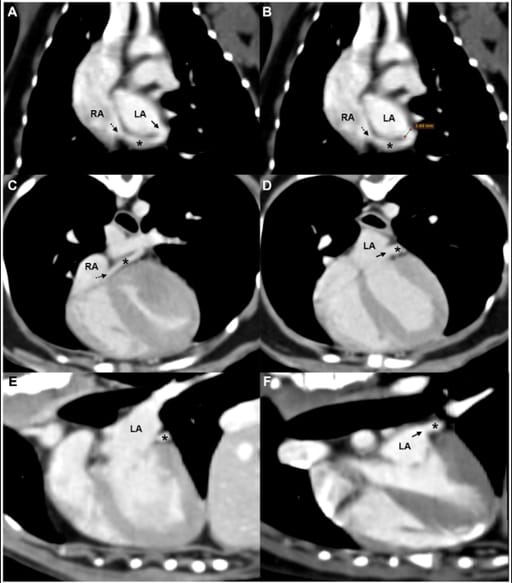
TTE showed a prominently visualized tubular structure compatible with the coronary sinus and continuous flow entering the right atrium (velocity ~0.5 m/s), without an atrial septal defect; Qp/Qs was 1.3, and a bubble study excluded a right-to-left shunt. CT confirmed a 3.6-mm partial defect between the mid-coronary sinus and the left atrium with similar contrast enhancement of both chambers, supporting a left-to-right shunt; no additional congenital anomalies were detected. Based on defect location and absence of persistent left superior vena cava, the lesion corresponds to a partially unroofed midportion (Type III by classic scheme; Type IIb by the Xie modification). Serial echocardiographic follow-up was recommended. WES highlighted four unique missense SNVs—CEP250, HHEX, CSPG4, CDK15—as candidate variants.
Limitations
This is a single-case report; the UCS defect was not directly visualized on TTE (advanced echocardiography or cardiac MRI might improve detection). ECG-gated CT was not performed, which can better depict small defects by reducing motion artefact. Genetic analysis was limited to one animal.
Conclusions
Asymptomatic UCS can occur in dogs and may be uncovered on screening when continuous right-atrial inflow from a markedly visualized coronary sinus is seen on TTE; CT angiography can confirm a small left-to-right shunt and precisely localize the defect. The case underscores the value of cross-sectional imaging, the need for surveillance for hemodynamic progression, and introduces candidate genes that warrant investigation in future cohorts.

Oblique reformatted CT images of the (A,B) dorsal, (C,D) axial, and (E,F) sagittal planes showing a partial defect between the coronary sinus and the LA. A tubular structure consistent with the coronary sinus (asterisk) courses along the posterior aspect of the LA and drains into the RA via the coronary sinus ostium (black dashed arrow). A partial defect (black solid arrow) in the septum between the coronary sinus and LA is visible in panels (A,D,F), with similar contrast enhancement noted between the two chambers. Measurement of the defect is shown in panel (B). LA, left atrium; RA, right atrium; *, coronary sinus.
How did we do? |
Disclaimer: The summary generated in this email was created by an AI large language model. Therefore errors may occur. Reading the article is the best way to understand the scholarly work. The figure presented here remains the property of the publisher or author and subject to the applicable copyright agreement. It is reproduced here as an educational work. If you have any questions or concerns about the work presented here, reply to this email.