- Veterinary View Box
- Posts
- Brush up on your elbow CT...
Brush up on your elbow CT...
Journal of Small Animal Practice, 2020
K. Baud, S. Griffin, F. Martinez-Taboada, N. J. Burton
Background
Elbow dysplasia is a common developmental disorder in dogs, particularly in Labrador Retrievers, with medial coronoid process disease (MCPD) being the most prevalent form. MCPD can result from abnormal bone development, subchondral disease, or incongruity of the elbow joint. While previous studies have examined elbow incongruity, differences in radioulnar congruity between radial incisure and apical medial coronoid process fragmentation remain unclear. This study aimed to compare elbow congruity between dogs with these two lesion types using computed tomography (CT).
Methods
A retrospective study was conducted using CT scans from Labrador Retrievers with elbow lameness diagnosed with MCPD. The study included 99 elbows from 66 dogs, divided into two groups: those with fragmentation at the radial incisure (56 elbows) and those with an apical medial coronoid process fragment (43 elbows). Radioulnar joint space was measured at multiple locations in the transverse, dorsal, and sagittal planes. Statistical analyses were performed to compare joint space measurements between the two groups.
Results
Overall, no significant differences in elbow congruity were found between the two groups except at a single measurement point in the transverse plane. At this location, dogs with radial incisure fragmentation had a narrower radioulnar joint space than those with apical fragmentation. This finding suggests that anatomic variation in the radial incisure group could create a fulcrum effect, leading to increased stress and predisposition to osteochondral damage at this site.
Limitations
This study used static CT scans, which do not account for dynamic changes in joint congruity under load. Additionally, the method of estimating joint space at the site of displaced fragments introduces potential measurement errors. The study did not include a control group of unaffected dogs, which could have provided additional context for the findings.
Conclusions
While no major differences in elbow congruity were found between radial incisure and apical medial coronoid process fragmentation, a subtle reduction in joint space at one measurement point suggests a potential mechanical contribution to radial incisure lesions. Further studies using dynamic imaging techniques are needed to confirm the clinical relevance of this finding and to better understand the role of elbow incongruity in MCPD.

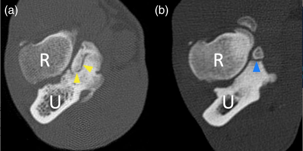
Transverse computer tomographic slice images demonstrating the two different types of medial coronoid process (MCP) fragmentation discussed in this study. (a) Fragmentation of the MCP along the radial incisure of the ulna (yellow arrow heads). (b) Fragmentation affecting the MCP at the apex (blue arrow head)
How did we do? |
Disclaimer: The summary generated in this email was created by an AI large language model. Therefore errors may occur. Reading the article is the best way to understand the scholarly work. The figure presented here remains the property of the publisher or author and subject to the applicable copyright agreement. It is reproduced here as an educational work. If you have any questions or concerns about the work presented here, reply to this email.