- Veterinary View Box
- Posts
- Cool VRU case report
Cool VRU case report
VRU 2025
Kari L. Means, Kekauilani Zukeran-Kerr, Kayla Le, Seng Wai Yap, Kelsey Brown, Lorelei Clarke
Background
Megaesophagus, a condition characterized by dilation and impaired motility of the esophagus, is rarely reported in small ruminants. Causes can include congenital defects, inflammation, or extraluminal compression. This study describes the first reported case of segmental megaesophagus in a goat due to esophageal stenosis caused by a transitional seventh cervical vertebra with supernumerary ribs.
Methods
Case Presentation:
-4-year-old Nigerian Dwarf wether with a three-year history of chronic regurgitation and cervical swelling.
-Progressive weight loss and lethargy despite normal appetite.
-Palpable firm mass along the ventral neck, eliciting discomfort and regurgitation upon manipulation.
Diagnostic Imaging:
-Radiographs: Identified a smoothly marginated, fusiform soft tissue mass in the cervical region, consistent with esophageal obstruction.
Computed Tomography (CT):
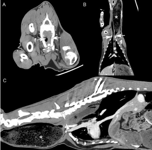
-Revealed a transitional seventh cervical vertebra with paired supernumerary ribs, causing severe esophageal stenosis at the thoracic inlet.
-The cervical esophagus was markedly dilated, containing heterogeneous fluid and gas, while the thoracic esophagus appeared normal.
Postmortem Examination:
-Confirmed compression of the esophagus by the abnormal ribs, resulting in severe esophageal dilation and chronic esophagitis.
Results
Pathophysiology:
-The supernumerary ribs were fused to the C7 vertebra, projecting ventromedially and compressing the esophagus at the thoracic inlet.
-This caused segmental stenosis, leading to proximal esophageal dilation (megaesophagus).
Prognosis and Outcome:
-Due to the chronicity and severity of esophageal dilation, surgical correction was deemed unlikely to restore normal function.
-The goat was humanely euthanized, and necropsy confirmed the CT findings.
Limitations
Single case report, limiting generalizability.
Lack of histopathologic analysis of esophageal muscle, which could provide insight into secondary neuromuscular involvement.
No long-term management attempted, as euthanasia was elected.
Conclusions
This is the first documented case of segmental megaesophagus in a goat due to extraluminal esophageal stenosis caused by a transitional C7 vertebra with supernumerary ribs. CT was essential in diagnosing the condition, highlighting the importance of advanced imaging in cases of chronic regurgitation and esophageal obstruction. Vertebral anomalies should be considered as a differential for extraluminal esophageal compression in ruminants, particularly when clinical signs appear progressively with age.

A, Transverse, (B) dorsal, and (C) sagittal reformatted images of postcontrast cervicothoracic CT demonstrating marked focal narrowing of the esophagus (white arrows) at the level of the paired C7 supernumerary ribs. Cranial to the site of compression, the esophagus is distended with intraluminal heterogeneous soft tissue attenuating material and gas (white asterisk), while the thoracic esophagus caudal to the site of compression contains fluid (black asterisk) and gas. CT images were obtained with slice thickness 1.25 mm, 120 kVp, 200 mA, soft tissue algorithm, viewed with window width 400 and window level 40.
How did we do? |
Disclaimer: The summary generated in this email was created by an AI large language model. Therefore errors may occur. Reading the article is the best way to understand the scholarly work. The figure presented here remains the property of the publisher or author and subject to the applicable copyright agreement. It is reproduced here as an educational work. If you have any questions or concerns about the work presented here, reply to this email.