- Veterinary View Box
- Posts
- CT and MRI Hallmarks of Canine Aortic Body Paragangliomas: Insights from 44 Cases
CT and MRI Hallmarks of Canine Aortic Body Paragangliomas: Insights from 44 Cases
Veterinary Radiology & Ultrasound (2025)
Obi Veterinary Education
January 16, 2026
Watko, M. A.; Mai, W.; McConnell, J. F.; Gomes, E.; Matz, B.; O’Brien, R. T.
Background
Aortic body paragangliomas are neuroendocrine tumors arising from chemoreceptor tissue at the heart base in dogs. These tumors are most commonly reported in brachycephalic breeds and may be incidental or associated with cardiorespiratory signs. Advanced imaging with computed tomography (CT) and magnetic resonance imaging (MRI) is increasingly used for diagnosis and staging; however, detailed descriptions of cross-sectional imaging characteristics in a large cohort of dogs have been limited.
Methods
This retrospective, multicenter study evaluated CT and MRI studies from 44 dogs with histologically or cytologically confirmed aortic body paragangliomas. Imaging examinations were reviewed for tumor location, size, margins, attenuation or signal characteristics, contrast enhancement patterns, vascular involvement, and evidence of local invasion or metastasis. Both pre- and post-contrast CT and MRI sequences were assessed when available.
Results
All tumors were located at the heart base, typically adjacent to the ascending aorta and pulmonary artery. On CT, masses were generally well-defined, soft-tissue attenuating, and showed moderate to marked contrast enhancement. Mineralization and cystic or necrotic regions were infrequently observed. On MRI, tumors were most commonly iso- to hypointense relative to muscle on T1-weighted images and heterogeneously hyperintense on T2-weighted images, with strong contrast enhancement. Vascular encasement or compression was commonly identified, while direct vascular invasion was less frequent. Metastatic disease was uncommon at the time of imaging.
Limitations
The study was limited by its retrospective design and variability in imaging protocols across institutions. Not all dogs underwent both CT and MRI, and histopathologic confirmation was not available for every lesion evaluated. Long-term outcome data were not consistently available for correlation with imaging findings.
Conclusions
Aortic body paragangliomas in dogs exhibit characteristic CT and MRI features, including strong contrast enhancement and close association with major heart base vessels. Cross-sectional imaging is valuable for tumor characterization, assessment of vascular involvement, and treatment planning. The imaging features described in this cohort may aid in differentiating aortic body paragangliomas from other heart base masses in dogs.
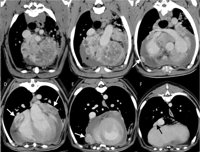
(A–F) Sequential postcontrast transverse CT images (from cranial to caudal) of an ABP with marked pericardial effusion (asterisk) and suspected pericardial (white arrow) and pleural (black arrow) metastases. WW = 352, WL = 62.
How did we do? |
Disclaimer: The summary generated in this email was created by an AI large language model. Therefore errors may occur. Reading the article is the best way to understand the scholarly work. The figure presented here remains the property of the publisher or author and subject to the applicable copyright agreement. It is reproduced here as an educational work. If you have any questions or concerns about the work presented here, reply to this email.