- Veterinary View Box
- Posts
- CT Imaging of Gastropexy in Dogs: Key Findings and Implications
CT Imaging of Gastropexy in Dogs: Key Findings and Implications
Journal of Small Animal Practice 2025
J. Einwaller, F. Llabres-Diaz, C. Jones, A. Caine
Background
Gastropexy is commonly performed to prevent gastric dilatation-volvulus (GDV) in dogs by creating a permanent adhesion between the stomach and body wall. Various techniques exist, each with different levels of adhesion and potential complications. This study aimed to describe the computed tomographic (CT) appearance of gastropexy sites, assess anatomic distortion post-surgery, and investigate potential imaging indicators of gastric functional abnormalities.
Methods
This retrospective study analyzed abdominal CT scans from 22 dogs that had undergone gastropexy. Dogs were categorized into anatomic (normal stomach positioning) and non-anatomic (abnormal stomach positioning) groups. CT images were evaluated for gastropexy site characteristics, gastric positioning, and signs of functional abnormalities such as gastric dilatation or gravel sign. Statistical comparisons were made between groups.
Results
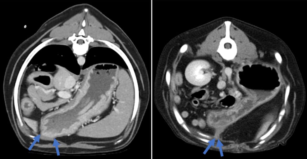
CT findings showed a median gastropexy site attenuation of 38.5 HU and a mild thickening of both the gastric and body wall musculature. Neovascularization was present in 65% of cases, and 32% exhibited marked gastric dilatation. A gravel sign, indicative of delayed gastric emptying, was observed in 73% of cases. The non-anatomic group had narrower gastropexy pedicles, shorter gastropexy lengths, and more acute gastric angles compared to the anatomic group. Despite anatomical distortion, gastrointestinal clinical signs were not always present.
Limitations
This study was limited by its retrospective nature, small sample size, and variation in surgical techniques. The exact impact of gastropexy on gastric motility remains unclear due to the lack of preoperative imaging and functional motility assessments. Variability in anesthesia and patient positioning may have influenced measurements.
Conclusions
CT imaging reveals significant variations in gastropexy sites, with some cases showing gastric distortion and potential indicators of functional abnormalities. While gastropexy may alter gastric anatomy, its impact on function requires further investigation. These findings provide a reference for radiologists assessing post-gastropexy changes and highlight the need for future studies on long-term outcomes and motility effects.

Post-contrast transverse CT images of the cranial abdomen at the level of the gastropexy site, displayed in a soft-tissue-window and illustrating a dog with a broad pedicle (left) and a dog with a narrow pedicle (right), marked by arrows.
How did we do? |
Disclaimer: The summary generated in this email was created by an AI large language model. Therefore errors may occur. Reading the article is the best way to understand the scholarly work. The figure presented here remains the property of the publisher or author and subject to the applicable copyright agreement. It is reproduced here as an educational work. If you have any questions or concerns about the work presented here, reply to this email.