- Veterinary View Box
- Posts
- CT Study Challenges Textbooks: Sciatic & Gluteal Lymph Nodes Found in Dogs
CT Study Challenges Textbooks: Sciatic & Gluteal Lymph Nodes Found in Dogs
VRU 2022
Riley J. Claude, Christopher P. Ober
Background
Traditional veterinary anatomy texts and the Nomina Anatomica Veterinaria state that dogs lack sciatic and gluteal lymph nodes, which are well described in other species. However, clinicians at the University of Minnesota observed structures resembling these lymph nodes in canine CT scans, especially in dogs with apocrine gland anal sac adenocarcinoma (AGASACA). The study aimed to determine the prevalence and characteristics of these lymph nodes in dogs and to assess their relevance for disease staging and treatment planning.
Methods
This retrospective anatomic study reviewed abdominopelvic CT scans from 121 dogs performed between September and December 2020. Two reviewers independently assessed images for the presence of sciatic and gluteal lymph nodes based on their expected anatomic locations in other species. Identified nodes were measured, and their contrast enhancement patterns described. Clinical records were reviewed to identify underlying disease processes, particularly neoplasia or lymphadenopathy.
Results
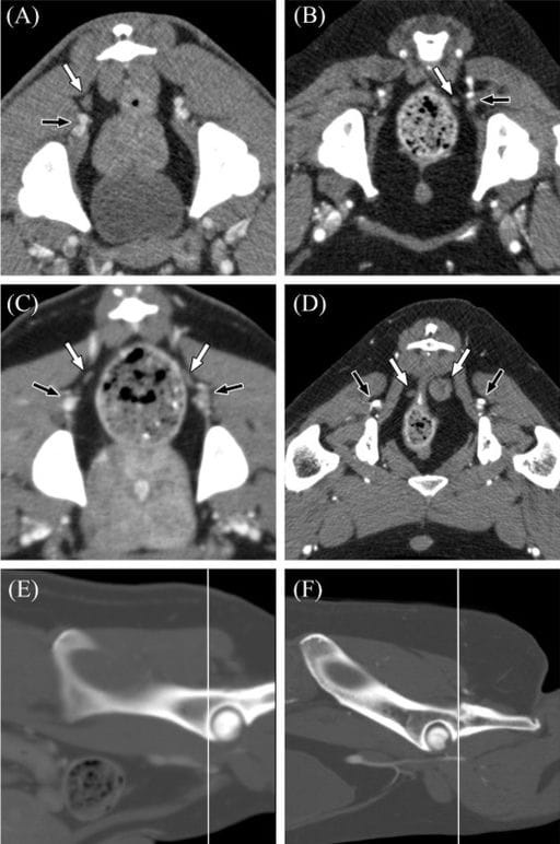
Sciatic lymph nodes were detected in 19 of 121 dogs (15.7%), with bilateral presence in 7 dogs and unilateral presence in 12. A single dog (0.8%) also exhibited bilateral gluteal lymph nodes, coinciding with prior AGASACA metastasis. Sciatic nodes were typically located near the caudal gluteal artery and vein, most often at the level of the cranial or mid-acetabulum. Their median long and short axes were 4.4 mm and 2.6 mm, respectively. Enhancement was mild to moderate in most nodes. While some nodes appeared normal, others were enlarged or heterogeneous, particularly in dogs with metastatic disease. Over half of affected dogs had concurrent lymphadenopathy elsewhere.
Limitations
No histopathological confirmation was performed to verify that the identified structures were lymph nodes. The study population included only dogs undergoing CT for clinical disease, introducing potential bias. Consequently, the normal size range and enhancement characteristics of these lymph nodes remain undetermined.
Conclusions
Contrary to long-held anatomical assumptions, sciatic and gluteal lymph nodes can be identified in a subset of dogs using CT imaging. Their recognition may hold clinical importance for staging and treatment of abdominopelvic neoplasia, such as AGASACA, where lymphatic spread affects prognosis and therapy planning. Further anatomical and histologic studies are warranted to confirm their presence and characterize their normal morphology.

Representative CT images of the positions of sciatic lymph nodes found in dogs. On transverse images, the patient's right is to the left of each image, and transverse images are displayed in a soft tissue window. A, Venous phase postcontrast transverse CT image of a canine pelvis at the level of the mid-acetabula demonstrating a right sciatic lymph node (white arrow) dorsal to the caudal gluteal artery and vein (black arrow). B, Early venous phase postcontrast transverse CT image of a canine pelvis at the level of the cranial to mid-acetabula demonstrating a left sciatic lymph node (white arrow) medial to the caudal gluteal artery and vein (black arrow). Note the poor enhancement of the caudal gluteal vein and other veins on this image due to the relatively early acquisition of the venous phase on this study. C, Venous phase postcontrast transverse CT image of a canine pelvis at the level of the cranial rims of the acetabula demonstrating bilateral sciatic lymph nodes (white arrows) dorsomedial to the respective caudal gluteal arteries and veins (black arrows). D, Venous phase postcontrast transverse CT image of a canine pelvis at the level of the caudal rims of the acetabula demonstrating bilateral sciatic lymph nodes (white arrows) dorsomedial to the respective caudal gluteal arteries and veins (black arrows). Because of the caudal position of these lymph nodes, the coccygeus muscle is seen between the lymph nodes and the respective caudal gluteal vessels. The left sciatic lymph node is the largest one identified in this study (14.9 × 14.5 mm). E, Sagittal reformed bone window CT image of the pelvis of the patient in (C) demonstrating the location of the transverse image (white line). F, Sagittal reformed bone window CT image of the pelvis of the patient in (D) demonstrating the location of the transverse image (white line)
How did we do? |
Disclaimer: The summary generated in this email was created by an AI large language model. Therefore errors may occur. Reading the article is the best way to understand the scholarly work. The figure presented here remains the property of the publisher or author and subject to the applicable copyright agreement. It is reproduced here as an educational work. If you have any questions or concerns about the work presented here, reply to this email.