- Veterinary View Box
- Posts
- CT Uncovers Hidden Lung Disease in Rabbits: Pulmonary Emphysema More Common Than Thought
CT Uncovers Hidden Lung Disease in Rabbits: Pulmonary Emphysema More Common Than Thought
VRU 2025
Athinodoros Athinodorou, Nicolas Israeliantz, Jenna Richardson, Dario Costanza, Jorge del Pozo, Tobias Schwarz
Background
Pulmonary emphysema (PE) is a rare but poorly understood respiratory condition in domestic rabbits, characterized by air trapping and destruction of lung tissue. While well-documented in humans, dogs, and cats, PE has previously been reported in rabbits only through isolated cases. This study aimed to describe the computed tomographic (CT) and clinical characteristics of PE in domestic rabbits and assess its prevalence, clinical correlations, and progression over time.
Methods
This retrospective, single-center case–control study analyzed 724 thoracic CT scans from 529 rabbits performed between 2017 and 2023 at the Royal (Dick) School of Veterinary Studies, University of Edinburgh. Fifty-nine rabbits with CT-confirmed PE and twenty-five PE-negative control rabbits were included. All CT scans were performed on conscious rabbits using standardized imaging protocols. Lesions were categorized by lobe involvement and emphysema type (diffuse, peripheral, bullous, mosaic), and attenuation values were measured in Hounsfield units (HU). Statistical analyses assessed associations between PE presence, lung attenuation, and clinical findings, with longitudinal follow-up for disease progression.
Results
PE was detected in 11.2% of rabbits (59/529). Affected rabbits had a mean age of 9 years, and the cranial lung lobes were most commonly involved. The median attenuation of emphysematous lung tissue (−905 HU) was significantly lower than nonemphysematous areas (−667 HU) and control lungs (−652 HU). Peripheral and bullous emphysema showed lower attenuation than diffuse types. No significant correlation was found between PE and lower respiratory tract clinical signs—44.1% of affected rabbits were asymptomatic. However, severe respiratory distress (e.g., open-mouth breathing, cyanosis) occurred in 10.2% of rabbits, all with advanced PE and poor prognosis. Secondary complications included rib fractures (5.1%) and pneumothorax (3.4%). In 15 rabbits with serial CTs, PE was progressive in 80% of cases.
Limitations
The study lacked histopathologic confirmation for all control subjects, leaving the possibility of undetected microscopic emphysema. The retrospective design and variable clinical data limited assessment of environmental or genetic risk factors. Additionally, some rabbits in the control group may have had subclinical pulmonary pathology not visible on CT.
Conclusions
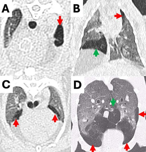
PE is a relatively common, progressive condition in older domestic rabbits and can occur without obvious respiratory signs. CT effectively identifies PE by revealing hypoattenuating lung regions with distinct patterns. Advanced disease may lead to complications such as pneumothorax or rib fractures. Clinicians should consider PE as a differential diagnosis in rabbits with respiratory distress and recognize its potential for progression even in asymptomatic cases.

CT images of emphysematous hypoattenuating lung tissue in rabbits. (A) Unilobar in the left cranial lung lobe (red arrow), and multilobar in three different rabbits in (B) the left cranial (red arrow) and right middle (green arrow) lung lobes, (C) both cranial lung lobes (arrows) and (D) both caudal (red arrows) and accessory (green arrow) lung lobes.
How did we do? |
Disclaimer: The summary generated in this email was created by an AI large language model. Therefore errors may occur. Reading the article is the best way to understand the scholarly work. The figure presented here remains the property of the publisher or author and subject to the applicable copyright agreement. It is reproduced here as an educational work. If you have any questions or concerns about the work presented here, reply to this email.