- Veterinary View Box
- Posts
- Discospondylitis part 2
Discospondylitis part 2
Journal of Veterinary Internal Medicine (JVIM) 2023
Cassie Van Hoof, Nicole A. Davis, Sheila Carrera-Justiz, Alisha D. Kahn, Steven De Decker, Nicholas J. Grapes, Michaela Beasley, John Du, Theresa E. Pancotto, Anna Suñol, Richard Shinn, Barry DeCicco, Erica Burkland, Harry Cridge
Background
Discospondylitis is an infection of the intervertebral disc and adjacent vertebral endplates, commonly caused by bacterial or fungal agents. Clinical signs vary from spinal pain to neurological deficits, and diagnosis is typically based on radiography, computed tomography (CT), or magnetic resonance imaging (MRI). However, the agreement between imaging modalities and risk factors for relapse or neurological deterioration remain unclear. This study aimed to:
-Describe the signalment, clinical features, imaging findings, treatment, and outcomes of dogs with discospondylitis.
-Assess diagnostic agreement between radiographs, CT, and MRI.
-Identify risk factors for relapse and progressive neurological deterioration.
Methods
Study Design: Multi-institutional retrospective study.
Study Population:
-386 dogs diagnosed with discospondylitis from 8 veterinary institutions (2010–2022).
-Signalment, clinical signs, imaging findings, diagnostic results, treatments, and outcomes were recorded.
Imaging Analysis:
-Radiographs, CT, and MRI were compared for lesion identification and location.
-Agreement between modalities was assessed using Cohen's kappa statistic.
Risk Factor Analysis:
-Evaluated history of trauma, steroid therapy, dental disease, neoplasia, and prior spinal surgery.
Results
Signalment and Clinical Signs:
-Male dogs were overrepresented (61%).
-The most affected site was L7-S1 (97/386 dogs, 26.8%).
-Spinal pain (63.5%) was the most common clinical sign, followed by paresis (30.6%).
-Fever was present in only 8% of cases, indicating it is not a reliable diagnostic marker.
Imaging Findings:
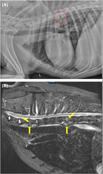
-Radiographs failed to detect discospondylitis in 33% of cases.
-CT and MRI detected additional lesions not seen on radiographs in 55 dogs (33%).
Agreement between modalities:
-Fair agreement between radiographs and CT (κ = 0.22).
-Poor agreement between radiographs and MRI (κ = 0.05).
-Good agreement between CT and MRI (κ = 0.72).
Microbiological Findings:
-Blood culture was positive in 27% of cases (most common: Staphylococcus spp.).
-Urine culture was positive in 28% of cases (most common: E. coli, Staphylococcus spp.).
-Brucella canis was detected in 6.6% of tested dogs.
-Fungal culture was positive in 21% of tested cases, with Aspergillus spp. most commonly identified.
Risk Factors for Poor Outcomes:
-History of trauma was associated with an increased risk of relapse (P = 0.01, OR = 9.0).
-Prior steroid therapy was associated with progressive neurological deterioration (P = 0.04, OR = 4.7).
Treatment and Outcomes:
-Empirical antibiotic therapy was most common (cephalexin, amoxicillin-clavulanate).
-Only 10.4% of dogs underwent surgery, typically for vertebral instability or abscess drainage.
-Clinical relapse occurred in 12% of dogs, with a median time to relapse of 12 months.
Limitations
Retrospective study design, with variability in diagnostic and treatment approaches.
Lack of standardization in follow-up imaging, leading to potential underestimation of disease progression.
No histopathologic confirmation of discospondylitis, relying on imaging and clinical criteria.
Conclusions
Radiographs have limited sensitivity for detecting discospondylitis, and MRI or CT should be pursued in suspected cases. Advanced imaging detects more lesions and improves localization accuracy. Male dogs and L7-S1 were most commonly affected, and Staphylococcus spp. was the predominant bacterial isolate. Trauma increased the risk of relapse, while prior steroid therapy was linked to neurological deterioration. This study highlights the need for advanced imaging in discospondylitis diagnosis and careful consideration of risk factors influencing prognosis.

(A and B) Representative images of a 9-year-old male neutered Labrador Retriever with discospondylitis. Advanced imaging such as MRI may detect more lesions than are evident on routine radiographs. (A) A left lateral radiographic projection reveals lysis of the caudal endplate of T5 and the cranial endplate of T6 (within red circle), along with spondylosis deformans at T5-T6 (^). (B) A midline STIR image of the cranial thoracic spine reveals irregularity and hyperintensity of the C7-T1 and T5-T6 intervertebral discs and adjacent cranial and caudal vertebral endplates (arrows).
How did we do? |
Disclaimer: The summary generated in this email was created by an AI large language model. Therefore errors may occur. Reading the article is the best way to understand the scholarly work. The figure presented here remains the property of the publisher or author and subject to the applicable copyright agreement. It is reproduced here as an educational work. If you have any questions or concerns about the work presented here, reply to this email.