- Veterinary View Box
- Posts
- Even in a Scotty, a bladder mass might not be a urothelial carcinoma...
Even in a Scotty, a bladder mass might not be a urothelial carcinoma...
Veterinary Radiology & Ultrasound, 2022
Hock Gan Heng, José A. Ramos-Vara, Christopher M. Fulkerson, Lindsey M. Fourez, Deborah W. Knapp
Background
Apex nodules in the urinary bladder were recently identified in Scottish Terriers undergoing bladder cancer screening. Given the high risk of urothelial carcinoma in this breed, distinguishing these nodules from neoplastic lesions is clinically significant. This study aimed to characterize the ultrasonographic features, prevalence, and clinical implications of apex nodules in Scottish Terriers.
Methods
A prospective, single-center case series was conducted at Purdue University. Scottish Terriers aged ≥6 years with no urinary tract disease were screened via ultrasonography and urinalysis every six months. The ultrasonographic characteristics of apex nodules, including location, margins, size, and echogenicity, were recorded. Cystoscopy and histopathology were performed in select cases. Follow-up examinations were conducted to assess changes in nodule appearance over time.
Results
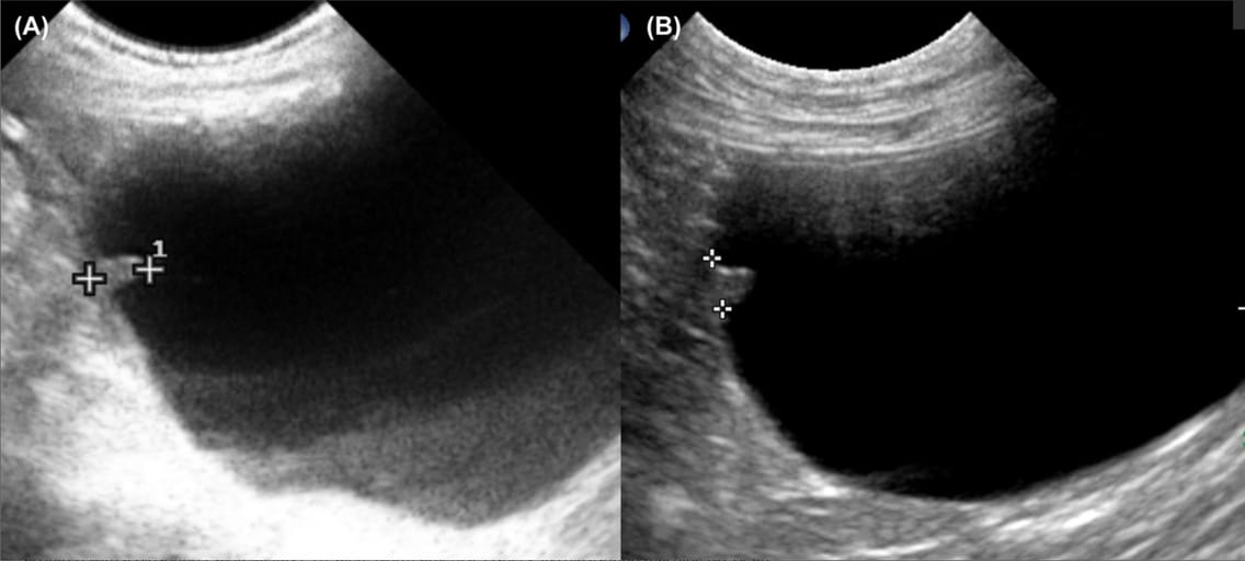
Apex nodules were identified in 6% (8/134) of Scottish Terriers. These nodules were single, well-defined, isoechoic to the bladder wall, and protruded into the lumen. Their base measured 2–4 mm, with a height of 4–6 mm. Five dogs underwent serial ultrasonographic evaluations (up to 3.5 years), with no observed changes in nodule size or morphology. Cystoscopic evaluation in three dogs revealed normal mucosal coverage, and histopathology in two dogs confirmed a benign fibrous structure with no neoplastic features. Three dogs later developed urothelial carcinoma at sites distant from the apex nodule, suggesting no direct association between the nodule and tumor development.
Limitations
The study had a small sample size, and histopathologic confirmation was available for only two cases. The imaging was performed primarily by a veterinary oncologist rather than a board-certified radiologist. Further studies with larger cohorts and detailed histological evaluation are needed to fully understand the origin and significance of apex nodules.
Conclusions
Apex nodules in the urinary bladder of Scottish Terriers appear to be benign, non-progressive structures that can mimic neoplastic lesions. These nodules should be considered in differential diagnoses when evaluating bladder abnormalities in this breed. Serial ultrasonography may help differentiate these benign structures from urothelial carcinoma, reducing unnecessary invasive procedures.

Longitudinal plane ultrasonographic images of two dogs. One small nodule was present at the apex of the urinary bladder in each dog (between the digital calipers). Note that the margins of the nodules were smooth and, they were protruding into the lumen. The apex nodules had an oblong shape (A) and triangular shape (B), respectively. For ultrasonography, the dog was placed in right lateral recumbency, the fur was clipped, and ultrasound gel was applied. B-mode ultrasound images of the urinary bladder distended with urine were acquired with a Biosound EsaoteMegas ES (Esaote North America, Fishers, IN) ultrasound machine (A), or an Aplio i800, Canon Medical Systems USA, Inc, (Tustin, California) ultrasound machine (B), and a 5–9 MHz microconvex array transducer in the longitudinal plane (A and B)
Disclaimer: The summary generated in this email was created by an AI large language model. Therefore errors may occur. Reading the article is the best way to understand the scholarly work. The figure presented here remains the property of the publisher or author and subject to the applicable copyright agreement. It is reproduced here as an educational work. If you have any questions or concerns about the work presented here, reply to this email.