- Veterinary View Box
- Posts
- Eyes That Retract: Dorsal Midbrain Lesions Cause Rare Nystagmus in Dogs
Eyes That Retract: Dorsal Midbrain Lesions Cause Rare Nystagmus in Dogs
Journal of Veterinary Internal Medicine, 2016
A.H. Crawford, E. Beltran, R. Lam, P.J. Kenny
Background
Convergence-retraction nystagmus is a distinct ocular movement disorder involving rhythmic convergence and retraction of the eyeballs, typically associated with Parinaud’s syndrome in humans due to dorsal midbrain lesions. This phenomenon had not previously been documented in veterinary literature. The study aimed to report and characterize convergence-retraction nystagmus in dogs and correlate it with dorsal midbrain pathology.
Methods
Three large-breed dogs presented with acute-onset vestibular signs underwent neurological examinations and advanced imaging. MRI was used to localize lesions, supplemented by CT and, in one case, cerebrospinal fluid analysis. Lesion characteristics were evaluated with T2-weighted, FLAIR, T1-weighted, and T2*-weighted fast field echo (FFE) MRI sequences. Clinical progression and outcomes were monitored over months.
Results
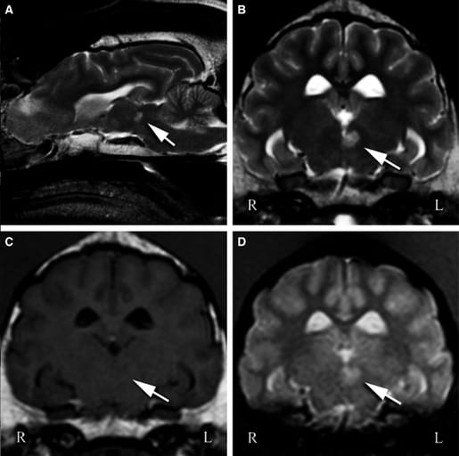
All three dogs demonstrated convergence-retraction nystagmus and vestibular signs (head tilt, ataxia). MRI revealed small, focal lesions in the dorsal midbrain near the rostral interstitial nuclei of the medial longitudinal fasciculus (RINMLF), consistent with cerebrovascular accidents. Lesions appeared hyperintense on T2W/FLAIR and non-enhancing post-contrast. No evidence of neoplasia or systemic inflammation was found. Two dogs recovered fully within weeks, and the third showed no recurrence for 34 months post-presentation. The abnormal eye movements were noted as key localizing signs for central vestibular disease.
Limitations
Definitive diagnosis was based on imaging without histopathologic confirmation. The small sample size and retrospective nature limited broader generalization. Some diagnostic procedures (e.g., CSF analysis) were incomplete in individual cases.
Conclusions
Convergence-retraction nystagmus is a highly specific sign of dorsal midbrain lesions in dogs and should be recognized as a distinct form of pathological eye movement. Its identification aids in neurolocalization and differentiation between central and peripheral vestibular syndromes. Awareness of this sign may improve diagnostic accuracy for midbrain cerebrovascular or structural disease in canine patients.

Case 3 T2-weighted sagittal image of the brain (A) and T2W (B), T1W (C), and FFE (D) transverse images at the level of the rostral midbrain revealed a round, well-demarcated lesion adjacent to the midline (left sided) and ventrolateral to the rostral part of the mesencephalic aqueduct. The lesion (indicated by the arrows) is hyperintense-to-normal-gray matter on T2W and FFE sequences, and is iso- to hypointense on T1W sequences.
How did we do? |
Disclaimer: The summary generated in this email was created by an AI large language model. Therefore errors may occur. Reading the article is the best way to understand the scholarly work. The figure presented here remains the property of the publisher or author and subject to the applicable copyright agreement. It is reproduced here as an educational work. If you have any questions or concerns about the work presented here, reply to this email.