- Veterinary View Box
- Posts
- Figures are beautiful.....
Figures are beautiful.....
PLOS One 2025
Samantha Miles, Charles McCauley, Mariano Carossino, Fabio Del Piero, Chin-Chi Liu, Lorrie Gaschen
Introduction
Manica flexoria tears are increasingly recognized as a cause of lameness in horses, but partial tears remain difficult to diagnose with conventional imaging. MRI is the best modality for evaluating soft tissues, yet the normal appearance of the manica flexoria on MRI has not been systematically described. The aim of this study was to characterize the normal MRI features and morphometry of the manica flexoria and to investigate differences between forelimbs and hindlimbs.
Materials and Methods
The study combined gross dissection and histology of two forelimbs and two hindlimbs from a sound horse with a retrospective MRI review of 18 non-lame limbs from 13 horses. MRI scans were obtained with a 1.5T magnet using proton density and T2-weighted sequences. Measurements of thickness were taken at four levels: proximal margin, proximal fourth, middle, and distal margin. Statistical analyses were performed using mixed ANOVA models with post-hoc comparisons.
Results
Grossly, the manica flexoria was thicker proximally in the forelimbs but thinner proximally in the hindlimbs, where it blended with overlying fascia and was overall shorter. Histology showed fibrous connective tissue layers, occasional fibrocartilaginous differentiation, and synovial lining, with the hindlimb structure being more uniform. On MRI, the manica flexoria was most often hyperintense to the superficial digital flexor tendon on proton density-weighted images and consistently isointense on T2-weighted images. A thin hyperintense line parallel to the dorsal margin was visible in all limbs. The proximal fourth was the thickest region in both limb types. In forelimbs, the medial aspect was thicker than the lateral, whereas in hindlimbs the lateral was thicker than the medial. No medial-lateral differences were noted at the distal margin.
Conclusion
The manica flexoria is thicker proximally than distally and shows distinct medial-lateral differences depending on whether it is located in the forelimb or hindlimb. These findings establish the first MRI-based reference values for the normal manica flexoria, which may improve the pre-operative diagnosis and prognostication of manica flexoria tears in horses.

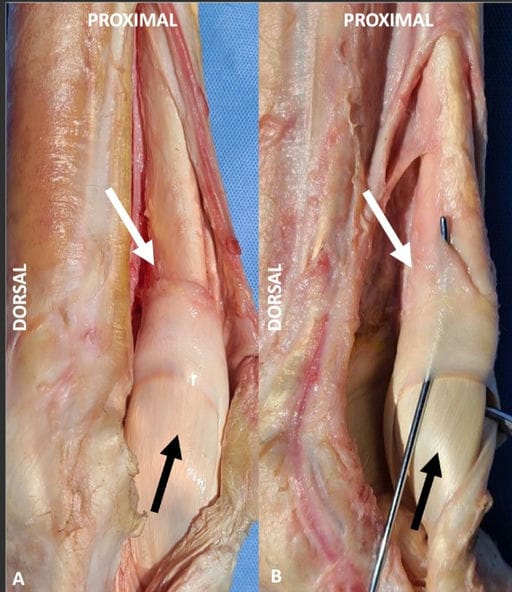
Gross anatomy of the manica flexoria and associated tendons.
Dissected images of a forelimb (A) and hindlimb (B) manica flexoria (white arrow), with the flexor tendons reflected, depicting the dorsal margin of the deep digital flexor tendon and the adjacent manica flexoria. The deep digital flexor tendon is seen passing through this structure (black arrow). Note the convex proximal margin and the angled medial and lateral distal margins of the manica flexoria. In the hindlimb, note that the proximal margin of the manica flexoria is ill-defined and blends into the fascia overlying the deep digital flexor tendon.
How did we do? |
Disclaimer: The summary generated in this email was created by an AI large language model. Therefore errors may occur. Reading the article is the best way to understand the scholarly work. The figure presented here remains the property of the publisher or author and subject to the applicable copyright agreement. It is reproduced here as an educational work. If you have any questions or concerns about the work presented here, reply to this email.