- Veterinary View Box
- Posts
- FIP in the thorax, what does it look like?
FIP in the thorax, what does it look like?
Journal of Feline Medicine and Surgery 2025
Kristin Repyak, Genna Atiee, Audrey Cook, Laura Bryan, Christine Gremillion
Background
Feline infectious peritonitis (FIP) is a fatal disease caused by feline coronavirus (FCoV), leading to systemic pyogranulomatous vasculitis. Diagnosis often relies on clinical signs, laboratory abnormalities, and imaging, particularly in cases lacking confirmatory histopathology. Although pleural effusion is a known feature of effusive FIP, detailed thoracic radiographic findings in FIP-affected cats remain underreported. This study aimed to describe thoracic radiographic abnormalities in cats with definitive or presumptive FIP.
Methods
A retrospective study was conducted on cats diagnosed with FIP between 2007 and 2022 at Texas A&M Veterinary Medical Teaching Hospital. Cases were included if they had a definitive FIP diagnosis (histopathology with immunohistochemistry) or a presumptive diagnosis (based on case review by two internists) and thoracic radiographs taken within seven days of clinical presentation. Radiographs were reviewed by a board-certified radiologist and a radiology resident for pulmonary, pleural, mediastinal, and cardiovascular abnormalities. Histopathologic findings were assessed in available cases.
Results
The study included 35 cats (18 with definitive FIP and 17 with presumptive FIP). Thoracic radiographs were abnormal in 91.4% (32/35) of cases. Pleural effusion was present in 37.1% (13/35), mostly bilateral. Pulmonary abnormalities were seen in 71.4% (25/35) of cats, with the most common pattern being unstructured interstitial (84%), followed by bronchial (44%), alveolar (40%), and nodular (12%). Sternal lymphadenopathy was present in 45.7% (16/35). Cardiomegaly was observed in 17.1% (6/35) and was linked to myocarditis, pericardial effusion, or high-output cardiac states. Pulmonary histopathology, available for 17 cats, showed pulmonary edema (94%), fibrinosuppurative pleuritis (76%), and histiocytic vasculitis (59%).
Limitations
The retrospective design and inclusion of both definitive and presumptive FIP cases introduce diagnostic uncertainty. Radiographic interpretation was subjective, despite consensus evaluation. The presence of pleural effusion may have confounded pulmonary pattern assessment. Additionally, not all cases underwent necropsy, limiting direct correlation between imaging and histopathology.
Conclusions
Cats with FIP frequently exhibit thoracic radiographic abnormalities, including pleural effusion, interstitial or bronchial lung patterns, lymphadenopathy, and cardiomegaly. While these findings are non-specific, FIP should be considered as a differential diagnosis in cats with compatible imaging and clinical signs. Further studies are needed to refine diagnostic criteria and assess imaging's prognostic value in FIP cases.

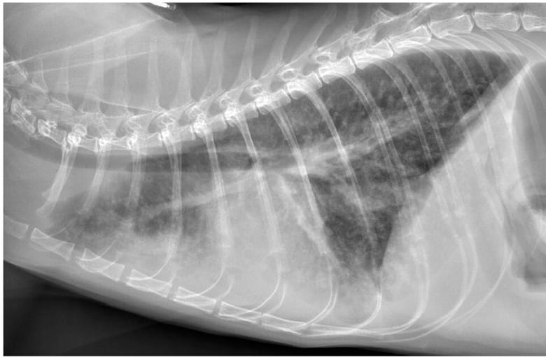
Right lateral thoracic radiograph (confirmed case, cat 15). Findings for this case included a severe diffuse interstitial pattern with numerous small nodules throughout all lung lobes, mild diffuse bronchial pattern, mild ventral alveolar pattern and mild pleural effusion. Pleural effusion silhouettes with the cranial and ventral margins of the cardiac silhouette, resulting in border effacement in those regions
How did we do? |
Disclaimer: The summary generated in this email was created by an AI large language model. Therefore errors may occur. Reading the article is the best way to understand the scholarly work. The figure presented here remains the property of the publisher or author and subject to the applicable copyright agreement. It is reproduced here as an educational work. If you have any questions or concerns about the work presented here, reply to this email.