- Veterinary View Box
- Posts
- First MRI-Confirmed Case of Wilson’s Disease in a Dog: A Diagnostic Breakthrough
First MRI-Confirmed Case of Wilson’s Disease in a Dog: A Diagnostic Breakthrough
VRU 2025
Natalie Durant, Matthew Paek, Wilfried Mai
Background
Wilson’s disease is an inherited disorder of copper metabolism caused by mutations in the ATP7B gene, leading to copper accumulation in the liver and brain. While well characterized in humans, the condition has not previously been confirmed in dogs. This case report describes the first documented MRI diagnosis of Wilson’s disease in a canine, detailing both neurological and hepatic involvement in a 3-year-old Dalmatian.
Methods
A 3-year-old castrated male Dalmatian presented with anorexia, vomiting, and lethargy that progressed to neurological deficits including ataxia, tetraparesis, and dull mentation. Diagnostic workup included hematology, serum biochemistry, abdominal ultrasonography, and brain MRI using a 1.5 T scanner. MRI sequences included T1-weighted, T2-weighted, T2-FLAIR, and post-contrast imaging. Liver biopsies were obtained for histopathology and quantitative copper analysis. The dog underwent copper chelation therapy with d-penicillamine and dietary modification, followed by repeat MRI evaluation after seven months.
Results
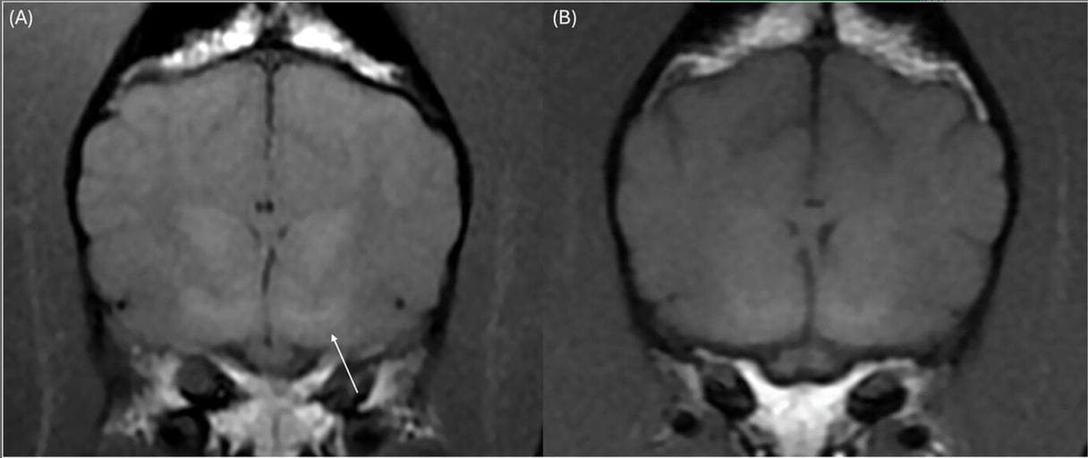
MRI revealed bilaterally symmetric, ill-defined T2- and T2-FLAIR hyperintensities in the thalami, geniculate bodies, red nuclei, and periaqueductal gray matter, with T1-hyperintensity in the lentiform nuclei and thalami—patterns consistent with metabolic or toxic encephalopathy. Liver histopathology demonstrated severe chronic copper-associated hepatitis, with a hepatic copper concentration of 3052 µg/g dry weight (normal < 400 µg/g). Following chelation therapy, clinical and neurological signs fully resolved, liver enzymes normalized, and follow-up MRI showed marked regression of previous lesions.
Limitations
As a single case study, the report cannot establish prevalence or genetic causation. No genetic testing for ATP7B mutations was performed. Pre-chelation MRI lacked a dorsal T2-weighted sequence, limiting the ability to identify the “face of the giant panda” sign typical in human Wilson’s disease. Although hyperammonemia and hepatic encephalopathy were ruled out, species-specific MRI variations may complicate direct comparison with human presentations.
Conclusions
This case documents the first MRI-confirmed diagnosis of Wilson’s disease in a dog, demonstrating parallel clinical, hepatic, and neuroimaging features to the human condition. The study emphasizes the value of MRI in detecting metabolic encephalopathies associated with copper storage diseases and highlights the potential for Dalmatians to serve as a preclinical model for Wilson’s disease. Prompt recognition and copper chelation can lead to complete neurological recovery and hepatic normalization.

(A) Bilaterally symmetric, ill-defined T1-hyperintensities in the globus pallidus (white arrow); (B) transverse T1W image showing bilaterally resolved lesions in the globus pallidus 7 months after copper chelation therapy was initiated.
How did we do? |
Disclaimer: The summary generated in this email was created by an AI large language model. Therefore errors may occur. Reading the article is the best way to understand the scholarly work. The figure presented here remains the property of the publisher or author and subject to the applicable copyright agreement. It is reproduced here as an educational work. If you have any questions or concerns about the work presented here, reply to this email.