- Veterinary View Box
- Posts
- For our European friends....
For our European friends....
Veterinary Radiology & Ultrasound, 2017
Mark E. Coia, Gawain Hammond, Daniel Chan, Randi Drees, David Walker, Kevin Murtagh, Janine Stone, Nicholas Bexfield, Lizzie Reeve, Jenny Helm
Background
Angiostrongylus vasorum is an emerging parasitic infection in dogs that primarily affects the cardiopulmonary system. Thoracic imaging is essential for diagnosing and monitoring the disease, yet descriptions of computed tomography (CT) findings in naturally infected dogs are limited. This multicenter retrospective study aimed to characterize thoracic CT abnormalities in dogs naturally infected with A. vasorum, assess whether common patterns exist, and propose standardized terminology for describing CT findings.
Methods
Medical records from nine UK-based referral centers were reviewed to identify dogs with confirmed A. vasorum infection that underwent thoracic CT between 2010 and 2015. Dogs were included if the diagnosis was confirmed via fecal smear, Baermann examination, bronchoalveolar lavage, ELISA, PCR, or serology, and if they had no concurrent disease that could cause similar pulmonary changes. CT scans were retrospectively evaluated, dividing lung lobes into pleural, subpleural, and peribronchovascular zones. Findings were categorized based on lung attenuation, lesion distribution, and severity.
Results
Eighteen dogs met the inclusion criteria. The predominant CT abnormality was increased lung attenuation due to ground-glass opacity or consolidation, often with a mosaic pattern. Lesions primarily affected the pleural and subpleural zones, with variable peribronchovascular involvement. Nine dogs (50%) had hyperattenuating nodules with ill-defined margins. Tracheobronchial lymphadenomegaly was a frequent finding. Bronchiectasis was observed in some cases, and mild pulmonary arterial dilation was noted in four dogs. No correlation was found between clinical severity and CT findings.
Limitations
The study's retrospective nature limited standardization of CT acquisition protocols. The natural disease course in dogs may differ from experimental infections, making direct comparisons difficult. Additionally, the study lacked follow-up imaging to assess changes post-treatment.
Conclusions
Thoracic CT findings in naturally infected dogs were consistent with a peripheral distribution of ground-glass opacity, consolidation, and nodules. These changes overlap with other pulmonary conditions, but A. vasorum should be considered a differential diagnosis in dogs with this pattern. This study provides a basis for standardized CT assessment and highlights the value of CT in diagnosing angiostrongylosis in clinical settings.

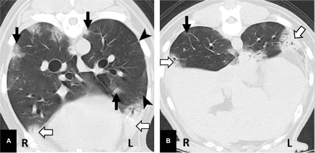
Transverse CT image of the thorax of a dog infected with A. vasorum obtained at the level of the right and left caudal lobes, and also includes the right accessory lung lobe (A). The caudal thorax is shown with the right and left caudal lung lobes given a score of 1 demonstrating mild parenchymal lesions (B). There are prominent parenchymal bands extending from the zone 1 into zone 2, with increased attenuation on the periphery of the lobe (black arrow head). Areas of patchy soft tissue attenuation resulting in effacement of the pulmonary vasculature, suggesting consolidation, are identifiable ventrally and in the caudal lung field; this is identifiable in both the left and right hemithorax (white arrow). Atelectasis (pertaining to cicatrization, compression or dependent) may be considered as a possible cause of the radiopathological sign. There is an ill-defined area of increased attenuation (GGO) within zones 2 and 3 (black arrow). There is a degree of bronchiolectasis identified in the left caudal lobe, seen in the peribronchovascular and subpleural zones. Window width (WW) 1400, window Level (WL) −500
How did we do? |
Disclaimer: The summary generated in this email was created by an AI large language model. Therefore errors may occur. Reading the article is the best way to understand the scholarly work. The figure presented here remains the property of the publisher or author and subject to the applicable copyright agreement. It is reproduced here as an educational work. If you have any questions or concerns about the work presented here, reply to this email.