- Veterinary View Box
- Posts
- GI ulcerations seems to be a think with intrahepatic portosystemic shunts....
GI ulcerations seems to be a think with intrahepatic portosystemic shunts....
Journal of the American Veterinary Medical Association (JAVMA), 2025
Erin A. Gibson, William T. N. Culp, Brian T. Hardy, Michelle A. Giuffrida, Ingrid M. Balsa, Phillip Mayhew, Stanley L. Marks
Background
Dogs with intrahepatic portosystemic shunts (IHPSS) frequently exhibit gastrointestinal (GI) abnormalities, including ulceration and hemorrhage. The role of percutaneous transvenous coil embolization (PTCE), a minimally invasive procedure used to treat IHPSS, in modifying GI mucosal lesions remains unclear. This study aimed to assess the prevalence and severity of GI mucosal lesions in dogs with IHPSS before and after PTCE using capsule endoscopy (CE).
Methods
A prospective clinical trial was conducted on ten client-owned dogs diagnosed with IHPSS via CT angiography. Capsule endoscopy was performed before PTCE and one month post-procedure to evaluate mucosal integrity. Data collected included mucosal lesion severity, serum biochemistry, and owner-reported clinical symptoms. Statistical analyses were conducted to compare findings before and after PTCE.
Results
Gastric and intestinal mucosal lesions were common in dogs with IHPSS, with 66.7% exhibiting gastric lesions and 62.5% showing intestinal lesions before PTCE. After PTCE, 80.0% had gastric lesions, while 40.0% had intestinal lesions. No significant difference was observed in mucosal lesion severity before and after PTCE. Despite this, owner-reported clinical symptoms improved significantly post-PTCE, particularly regarding malaise and GI signs.
Limitations
The study was limited by its small sample size and short follow-up period. Some capsule endoscopy studies were incomplete, potentially underestimating lesion prevalence. The clinical significance of subclinical mucosal lesions remains uncertain, and long-term effects of PTCE on GI health require further investigation.
Conclusions
Subclinical GI mucosal lesions are common in dogs with IHPSS, regardless of PTCE treatment. While PTCE significantly improved clinical signs, it did not influence the prevalence or severity of mucosal lesions in the short term. These findings suggest that dogs with IHPSS may require ongoing GI management independent of shunt attenuation. Future research should explore the long-term clinical impact of these lesions and the potential benefits of GI-protective therapies.

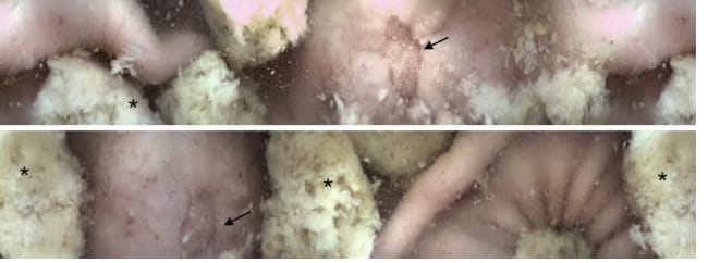
Capsule endoscopy image obtained in the stomach of dog 10 one month following percutaneous transvenous coil embolization (PTCE). A resolving ulcer (grade 6 mucosal lesion; black arrows) is observed in the gastric wall. Note the gastric fluid and concurrent ingesta (asterisks) obscuring some of the gastric wall.
How did we do? |
Disclaimer: The summary generated in this email was created by an AI large language model. Therefore errors may occur. Reading the article is the best way to understand the scholarly work. The figure presented here remains the property of the publisher or author and subject to the applicable copyright agreement. It is reproduced here as an educational work. If you have any questions or concerns about the work presented here, reply to this email.