- Veterinary View Box
- Posts
- High SLN Detection in Canine Oral Tumors with CT & Methylene Blue
High SLN Detection in Canine Oral Tumors with CT & Methylene Blue
JAVMA 2025
Sohee Bae, Charly Mckenna, Stephanie Goldschmidt, Owen T. Skinner, Judith Bertran, Debbie Reynolds, Michelle L. Oblak
Background
Sentinel lymph node (SLN) mapping improves cancer staging by identifying the primary lymph node likely to harbor metastases. This approach reduces unnecessary lymphadenectomy and associated morbidity. In veterinary medicine, reliable SLN detection remains challenging, especially in canine head and neck cancers due to complex lymphatic drainage and variability. CT lymphangiography (CTL) and intraoperative methylene blue (IOL-MB) are emerging alternatives to radioactive tracer-based techniques, but comparative data are limited.
Methods
This was a prospective study of 38 client-owned dogs with oral neoplasms (macroscopic or incompletely excised malignant tumors). Dogs underwent preoperative CTL with iodinated contrast and intraoperative SLN mapping with peritumoral methylene blue injection. Bilateral mandibular and medial retropharyngeal lymph nodes were surgically removed and analyzed. Agreement between CTL and IOL-MB was assessed using Cohen’s kappa (κ), and detection accuracy was evaluated based on histopathological confirmation of metastases.
Results
The combined CTL and IOL-MB approach achieved a 97.4% SLN detection rate. Moderate agreement was observed between the two modalities (76.8%, κ = 0.485). All 6 metastatic LNs (from 4 dogs) were identified using CTL, and 5 of 6 with IOL-MB. Nine dogs had discrepancies between the two mapping techniques, including one where IOL-MB failed to detect a metastatic node. Bilateral SLNs and contralateral drainage patterns were noted in several cases, emphasizing the anatomical variability.
Limitations
The study had a small sample size and low rate of lymph node metastasis (2.7%), partly due to exclusion of cases with overt nodal involvement. Different institutions used varying contrast agents, and there was no centralized pathology review. The timing of IOL-MB administration and node removal was variable and may have influenced SLN identification. Prior excisional biopsies and patient body size may have also affected lymphatic drainage.
Conclusions
CTL and IOL-MB provide a high SLN detection rate with moderate inter-modality agreement. The techniques are complementary and should be used together, especially in the absence of clinically evident nodal metastasis. Anatomical variability and lymphatic flow alterations from metastasis can influence SLN identification, underlining the importance of using multiple modalities and evaluating all suspicious nodes. Further research is needed in larger populations with higher metastatic burden to validate and refine SLN mapping protocols.

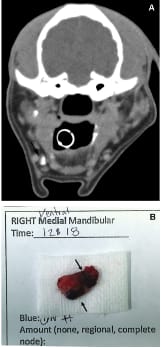
Sentinel lymph node (SLN) mapping techniques. A—Transverse indirect CT lymphangiography image obtained 3 minutes after completing the peritumoral injection. The LN was defined as an SLN when it was contrasted in this technique. A ventromedial mandibular LN was contrasted in this dog and was considered an SLN (black arrow). B—Intraoperative methylene blue lymphangiography. The LN that was stained ≥ 1 was considered to be the SLN. The right medioventral mandibular LN was defined as a SLN.
How did we do? |
Disclaimer: The summary generated in this email was created by an AI large language model. Therefore errors may occur. Reading the article is the best way to understand the scholarly work. The figure presented here remains the property of the publisher or author and subject to the applicable copyright agreement. It is reproduced here as an educational work. If you have any questions or concerns about the work presented here, reply to this email.