- Veterinary View Box
- Posts
- How can we see those feline carpal ligaments?
How can we see those feline carpal ligaments?
BMC Veterinary Research 2022
Rachel M. Basa, Kenneth A. Johnson, Juan M. Podadera
Background
Feline carpal injuries are typically diagnosed using radiography and computed tomography (CT), but these modalities do not allow direct visualization of carpal ligaments. Magnetic resonance imaging (MRI) and arthrography are used in humans to enhance ligament visualization, but their utility in cats has not been well studied. This study aimed to evaluate the effectiveness of CT arthrography (CTA) and MR arthrography (MRA) in visualizing feline carpal ligaments, comparing them to conventional CT and MRI. A secondary objective was to describe a protocol for intra-articular contrast injection in the feline carpus.
Methods
Study Design: Ex-vivo cadaveric study.
Specimens:
-Five paired feline cadaveric forelimbs from skeletally mature Domestic Short Hair cats.
-Three additional canine antebrachii used for contrast pilot study.
Contrast Protocol:
-A mixture of gadolinium (0.05 mmol/mL) and iohexol (150 mgI/mL) was injected into the antebrachiocarpal and middle carpal joints under fluoroscopic guidance.
-Volume used: 0.8 mL for antebrachiocarpal joint, 0.5 mL for middle carpal joint.
Imaging Techniques:
-Plain CT and CTA performed using a 16-slice Philips Brilliance CT scanner.
-MRI and MRA performed using a 3.0 Tesla GE Healthcare scanner, with T1-weighted sequences (pre- and post-contrast) and fat suppression.
-Images were analyzed in sagittal, transverse, and dorsal planes by a board-certified radiologist and surgeon.
Scoring System:
Ligament visibility was graded on a 0 to 3 scale, where:
-0 = not visible,
-1 = partially visible,
-2 = visible but poorly demarcated,
-3 = fully visible and well demarcated.
Results
CT vs. CTA:
-Plain CT did not allow identification of any ligaments.
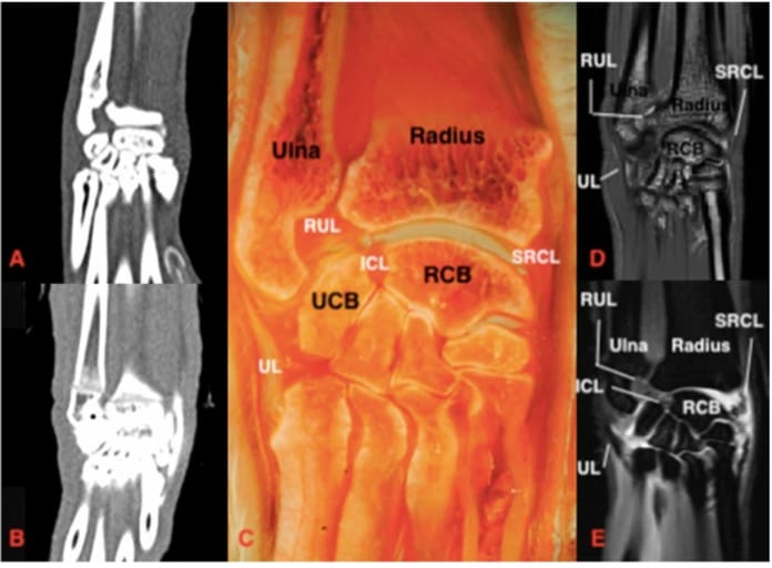
-CTA enabled visualization of intra-articular ligaments and some extra-articular ligaments (e.g., short radial and short ulnar collateral ligaments) when joint distension was adequate.
MRI vs. MRA:
-MRI was superior to CTA for identifying carpal ligaments.
-MRA further improved ligament visualization, especially for the dorsal radiocarpal, accessorioulnocarpal, accessorioquartile, and short collateral ligaments.
-Palmar radiocarpal and ulnocarpal ligaments were best visualized on MRA in the sagittal and dorsal planes.
Joint Communication Findings:
-Antebrachiocarpal and middle carpal joints did not communicate.
-Contrast flowed dorsodistally within the antebrachiocarpal joint, suggesting that the middle carpal joint should be injected first to avoid leakage.
Limitations
Cadaveric study design—results may not fully translate to live animals.
Freeze-thaw cycles may have affected tissue properties and imaging clarity.
Small sample size (n = 5) limits statistical analysis.
Subjective evaluation of ligament visibility, with potential observer bias.
Conclusions
MRA provided the best visualization of feline carpal ligaments, outperforming both CTA and conventional MRI. CTA may still be useful when MRI is unavailable, particularly in cases with concurrent carpal bone fractures. These findings support the use of arthrography to enhance carpal ligament evaluation in feline patients with suspected ligamentous injury. Further studies in live cats are needed to validate these results for clinical applications

Dorsal section of the feline carpus. Proximal is at the top of the image and lateral is to the left. A Plain CT, soft tissue window B) CTA, soft tissue window C) Gross plastinated section D) T1W E) T1W FS post contrast. Note that the silhouette of the SRCL can be seen in B) post contrast injection. There is improved visibility of the SRCL in the post contrast MRI (E compared to D). RCB: radial carpal bone, UCB: ulnar carpal bone, RUL: radioulnar ligament, UL: ulnaris lateralis, ICL: radioulnar intercarpal ligament, SRCL: short radial collateral ligament
How did we do? |
Disclaimer: The summary generated in this email was created by an AI large language model. Therefore errors may occur. Reading the article is the best way to understand the scholarly work. The figure presented here remains the property of the publisher or author and subject to the applicable copyright agreement. It is reproduced here as an educational work. If you have any questions or concerns about the work presented here, reply to this email.