- Veterinary View Box
- Posts
- How much contrast should you inject in the fetlock?
How much contrast should you inject in the fetlock?
Research in Veterinary Science 2025
Cartilage Defect Identification on Computed Tomography Arthrography in Equine Fetlock: Ex-Vivo Study
Irene Nocera, Chiara Di Franco, Elisa Marcucci, Caterina Puccinelli, Giulia Sala, Micaela Sgorbini, Simonetta Citi
Background
Computed tomography arthrography (CTA) is a valuable diagnostic imaging technique for detecting cartilage defects in equine joints. However, the volume of intra-articular contrast medium (CM) required for optimal visualization has not been standardized and is often based on operator experience. This study aimed to determine the minimum CM volume sufficient for detecting iatrogenic cartilage defects in the equine fetlock joint using ex-vivo specimens.
Methods
The study was conducted on 32 distal limbs from adult horses obtained from a slaughterhouse. Full-thickness cartilage defects were arthroscopically created at five predetermined locations in the fetlock joint. A series of CTA scans were performed on each joint, beginning with a precontrast scan, followed by scans with increasing CM volumes ranging from 2.5 mL to 40 mL. The presence and volume of cartilage defects were assessed using CTA and compared to macroscopic measurements. Statistical analyses included chi-square tests for defect detection and Spearman’s correlation for volumetric comparison.
Results
Cartilage defects were detected in only 6% of cases (10/160) in precontrast scans (0 mL CM), but all defects were visible when a minimum of 20 mL CM was used. A volume of 5 mL was sufficient to detect 96% of defects, particularly in certain joint regions. A weak correlation was observed between defect volume measurements obtained from CTA and macroscopic evaluation.
Limitations
The study used ex-vivo specimens, which may not fully replicate in-vivo conditions. Freezing and thawing of the joints could have affected tissue integrity and joint fluid dynamics, potentially impacting CM distribution. The study also relied on a single observer for image analysis, which may introduce bias in defect measurements.
Conclusions
A CM volume of 20 mL was sufficient to detect all cartilage defects in the equine fetlock joint using CTA, supporting the standardization of contrast volume for clinical use. The findings suggest that optimizing CM use could improve diagnostic consistency while minimizing potential side effects associated with excessive CM administration.

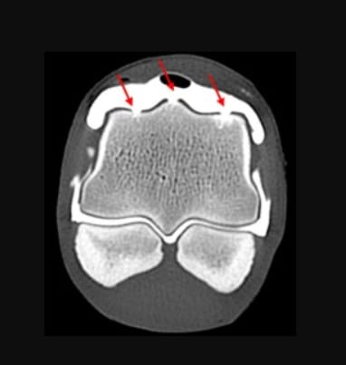
CTA scan, at the volume of iodinated contrast of 20 ml, displayed in a transverse plane generated via multiplanar reconstruction. Note the site of the defects in the metacarpal medial and lateral condyles, and sagittal ridge were indicated by the red arrows. Images were created using the MPR function in a bone window (WL: 553 WW: 2613) from bone kernel reconstruction.
How did we do? |
Disclaimer: The summary generated in this email was created by an AI large language model. Therefore errors may occur. Reading the article is the best way to understand the scholarly work. The figure presented here remains the property of the publisher or author and subject to the applicable copyright agreement. It is reproduced here as an educational work. If you have any questions or concerns about the work presented here, reply to this email.