- Veterinary View Box
- Posts
- Intramedullary Gas on CT: Recognizing Emphysematous Osteomyelitis in Dogs Before It’s Too Late
Intramedullary Gas on CT: Recognizing Emphysematous Osteomyelitis in Dogs Before It’s Too Late
Veterinary Radiology & Ultrasound (2025)
Obi Veterinary Education
January 11, 2026
Anastasia M. McHaney; Katherine A. Weber; Erica Chávez-Peón Berle; Michelle Riehm; William H. Whitehouse; Seng Wai Yap; Nicolette Cassel
Background
Osteomyelitis is a relatively common condition in veterinary patients; however, emphysematous osteomyelitis, defined by the presence of intramedullary gas, is exceedingly rare in both veterinary and human medicine. In human patients, this entity is associated with significant mortality and is often linked to systemic comorbidities. Prior to this report, emphysematous osteomyelitis had been documented only once in dogs. The study aimed to describe the imaging characteristics, clinical progression, and outcomes of two canine cases and to contextualize these findings within the existing veterinary and human literature.
Methods
This retrospective descriptive case series evaluated two young dogs presenting to tertiary veterinary centers with acute inflammatory and infectious clinical signs. Inclusion criteria required complete medical records, CT-confirmed intramedullary gas, and positive bacterial cultures from adjacent soft tissues. Diagnostic evaluation included radiography, ultrasonography, and computed tomography, with imaging interpreted by diagnostic imaging residents and board-certified radiologists. Clinical data, imaging findings, culture results, treatment, and follow-up outcomes were reviewed.
Results
Both dogs demonstrated multiple small, irregularly marginated intramedullary gas foci within the femur on CT, producing a “pumice stone” appearance characteristic of emphysematous osteomyelitis. Peri-femoral abscessation was present in both cases, and bacterial cultures were positive (Clostridium haemolyticum in one case and beta-hemolytic Streptococcus spp. in the other). One dog responded favorably to prolonged antimicrobial therapy, with progressive imaging changes consistent with chronic osteomyelitis followed by near-complete osseous remodeling and resolution of intramedullary gas. The second dog experienced rapid systemic deterioration with suspected septic thromboembolic disease and was euthanized despite aggressive medical and surgical intervention.
Limitations
The study was limited by its retrospective design and the extremely small sample size inherent to the rarity of the condition. Lack of standardized treatment protocols and limited long-term outcome data restrict broader prognostic conclusions. Additionally, definitive routes of infection could not be established in either case.
Conclusions
Emphysematous osteomyelitis represents a rare but severe form of osteomyelitis in dogs, associated with a guarded prognosis and potential for rapid clinical decline. CT is the imaging modality of choice for diagnosis, as the presence of multiple small intramedullary gas foci without a history of trauma or surgery is considered pathognomonic. Early recognition and aggressive, prolonged antimicrobial therapy may improve outcomes. Increased awareness and early CT utilization may lead to earlier diagnosis and better characterization of this underrecognized condition in veterinary medicine.
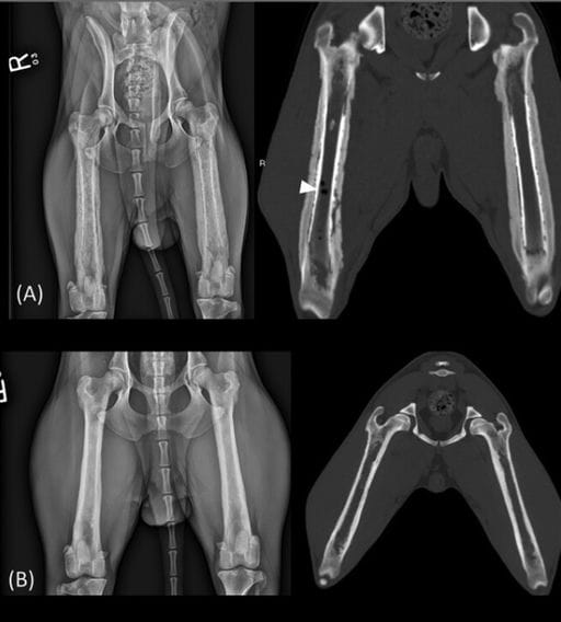
Follow-up extended ventrodorsal pelvic radiographs and CT in bone window, reformatted into dorsal plane, of Case 1. (A) Four-week recheck. Note the exuberant, circumferential, palisading to thick and smooth periosteal reaction, with concurrent cortical lysis and subperiosteal scalloping; linear, hypoattenuating subperiosteal regions parallel to the femoral cortices; and hyperattenuating, sclerotic intramedullary regions that replaced previously identified intramedullary gas bubbles—all consistent with chronic remodeling secondary to osteomyelitis. Also note the persistence of the intramedullary gas bubbles seen on CT (white arrowhead). (B) On the 4-month recheck, note the near complete and marked improvement in opacity and thickness of the femoral cortices, with absence of intramedullary gas.
How did we do? |
Disclaimer: The summary generated in this email was created by an AI large language model. Therefore errors may occur. Reading the article is the best way to understand the scholarly work. The figure presented here remains the property of the publisher or author and subject to the applicable copyright agreement. It is reproduced here as an educational work. If you have any questions or concerns about the work presented here, reply to this email.