- Veterinary View Box
- Posts
- Is the CSF flow-void sign influenced by heart rate or blood pressure in small dogs?
Is the CSF flow-void sign influenced by heart rate or blood pressure in small dogs?
Vet Radiol Ultrasound. 2009
Obi Veterinary Education
January 23, 2026
Sean R. Freer, Peter V. Scrivani, Hollis N. Erb
Background
The cerebrospinal fluid (CSF) signal-void sign on T2-weighted MRI reflects CSF signal loss and is commonly observed in small-breed dogs with hydrocephalus and syringomyelia. In humans, this sign is attributed to high-velocity or turbulent CSF flow related to arterial pulsations and reduced intracranial compliance. Cardiovascular variables such as heart rate, blood pressure, and end-tidal CO₂ may influence CSF flow or intracranial pressure, particularly under anesthesia. This study aimed to determine whether such cardiopulmonary variables are associated with the presence of the CSF signal-void sign in small-breed dogs.
Methods
A retrospective study was performed on 53 anesthetized small-breed dogs (<15 kg) that underwent spin-echo, T2-weighted MRI of the brain using a standardized protocol. The CSF signal-void sign in the mesencephalic aqueduct was assessed subjectively and dogs were classified as either having or not having the sign. Cardiopulmonary variables recorded during image acquisition included heart rate, systolic, mean, diastolic, and pulse blood pressure, and end-tidal CO₂. Statistical analyses included tests for normality, Student’s t-tests for group comparisons, and Pearson’s correlation analyses.
Results
A CSF signal-void sign was identified in 19 of 53 dogs (36%). No statistically significant differences were detected between dogs with and without the CSF signal-void sign for heart rate, any blood pressure measure, pulse pressure, or end-tidal CO₂. Correlation analyses showed expected relationships among blood pressure variables but no meaningful associations between end-tidal CO₂ and other measures. Statistical power for detecting group differences was low for all variables assessed.
Limitations
The primary limitation was small sample size, resulting in low statistical power and a risk of Type II error. The retrospective design and lack of standardized anesthetic protocols introduced variability in cardiopulmonary measurements. Additionally, only routinely recorded variables were assessed, and image acquisition parameters, while relatively standardized, may still influence detection of the CSF signal-void sign.
Conclusions
No association was identified between cardiopulmonary variables and the presence of a CSF signal-void sign in small-breed dogs under anesthesia. However, due to limited statistical power, the absence of detected differences does not exclude a true association. If such a relationship exists, it is likely weak. Further research is needed to clarify the pathophysiology and clinical relevance of the CSF signal-void sign in dogs.
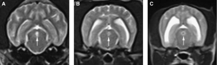
Transverse, spin echo T2-weighted MR images of the mesencephalon of three different small-breed dogs depicting no cerebrospinal fluid (CSF)signal-void sign (A), an intermediate-intensity CSF signal-void sign (B), and a pronounced CSF signal-void sign (C) in the mesencephalic aqueduct (arrow). Forthe statistical analyses, both the intermediate CSF signal-void sign and pronounced CSF signal-void sign were considered as definitively detected
How did we do? |
Disclaimer: The summary generated in this email was created by an AI large language model. Therefore errors may occur. Reading the article is the best way to understand the scholarly work. The figure presented here remains the property of the publisher or author and subject to the applicable copyright agreement. It is reproduced here as an educational work. If you have any questions or concerns about the work presented here, reply to this email.