- Veterinary View Box
- Posts
- Is there an association between lumbosacral transitional vertebra (LTV) and canine hip dysplasia?
Is there an association between lumbosacral transitional vertebra (LTV) and canine hip dysplasia?
Acta Veterinaria Scandinavica 2025
Jon Andre Berg, Bente Kristin Sævik, Frode Lingaas, Catrine Trangerud
Background
A lumbosacral transitional vertebra (LTV) is a congenital vertebral anomaly that exhibits characteristics of both lumbar and sacral vertebrae. It has been associated with canine hip dysplasia (CHD) and cauda equina syndrome (CES), particularly in German Shepherds. Prior studies suggest that asymmetrical LTVs may contribute to asymmetrical CHD grades due to altered pelvic biomechanics. This study aimed to evaluate the relationship between asymmetrical LTVs and pelvic morphology using ventrodorsal (VD) radiographs and assess whether LTV-related pelvic rotations contribute to asymmetrical CHD development.
Methods
Study Design: Retrospective cross-sectional study.
Study Population:
-13,950 dogs from 14 breeds were included from the Norwegian Kennel Club (NKK) radiographic screening program for CHD between 2014 and 2022.
-2579 dogs (18.5%) had LTV, classified as symmetrical (78.6%) or asymmetrical (21.4%).
Radiographic Classification:
-LTVs were categorized into three types based on vertebral morphology.
-Asymmetrical CHD was identified in 12.4% of dogs, and the prevalence of asymmetrical LTV among these cases was 39.7%.
Measurements and Analysis:
-Pelvic rotation was assessed in vertical and longitudinal axes.
-The sacroiliac joint (SIJ) length and its positional shift were measured relative to the hip joint.
-Statistical models were used to evaluate associations between LTV morphology, pelvic rotation, and CHD grading.
Results
Asymmetrical LTV was associated with pelvic counter-rotation (P < 0.001), suggesting a compensatory mechanism to straighten the lower back.
The sacroiliac joint length was significantly reduced on the shorter side of an asymmetric LTV, causing a caudal shift of the SIJ and a reduced distance to the hip joint (P < 0.001).
Rotation of an asymmetrical LTV in the longitudinal axis was linked to contralateral pelvic rotation (P < 0.001), potentially influencing CHD development.
An asymmetrical CHD grade was more likely in dogs with an asymmetrical LTV (P < 0.001), indicating a biomechanical link between LTV and hip joint malformation.
Limitations
Retrospective design with reliance on existing radiographs.
No direct biomechanical testing, making causal relationships speculative.
Variability in radiographic positioning may have introduced minor measurement errors.
Conclusions
Asymmetrical LTVs are associated with altered pelvic rotation and sacroiliac joint positioning, which may contribute to asymmetrical CHD development. The findings support the theory that counter-rotation between the pelvis and LTV segment helps maintain spinal alignment but may also predispose dogs to hip joint malformations and cauda equina syndrome. Future research should explore biomechanical implications and genetic predisposition for LTV-associated orthopedic disorders.

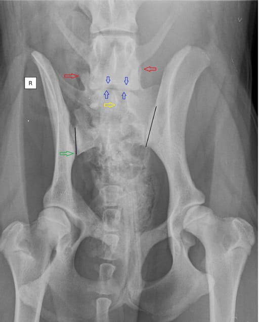
Ventrodorsal radiography with extended hips reveals an asymmetric lumbosacral transitional vertebra (LTV). The lumbosacral transitional vertebra
(LTV) segment exhibits a sacral-like transverse process with a broad contact area on the left. The rotation of the LTV segment is evident on both the verticalmand longitudinal axes, leaning towards the left. This is observed through the spinous process (yellow arrow), cranial articular facets (red arrows), and cranial and caudal endplates compared to the last normal lumbar vertebra (blue arrows). The pelvis demonstrates longitudinal axis rotation, noticeable in the wider left iliac wing and the asymmetric appearance of the obturator foramen. Additionally, a counterclockwise vertical rotation of the pelvis and sacrum is observed and a caudal displacement of the sacroiliac joint (green arrow). This displacement shortens the distance from the sacroiliac joint to the hip joint on the right side. Moreover, inadequate coverage of the left acetabulum leads to subluxation of the left hip joint. Axial malpositioning of a normal dog during the radiographic examination results in apparent rotation of the pelvis and the caudal lumbar spine in the same direction. However, if the direction or degree of the rotation, or both together, between the pelvis and lumbar spine are different, an inherent malposition should be considered [8]. The black lines demonstrate the measurement of the sacroiliac joint length. (The radiograph is for illustration purposes and not from the NKK database)
How did we do? |
Disclaimer: The summary generated in this email was created by an AI large language model. Therefore errors may occur. Reading the article is the best way to understand the scholarly work. The figure presented here remains the property of the publisher or author and subject to the applicable copyright agreement. It is reproduced here as an educational work. If you have any questions or concerns about the work presented here, reply to this email.