- Veterinary View Box
- Posts
- Is this a possible sequela of kennel cough?
KIMBERLY B. WINTERS, AMY S. TIDWELL, ELIZABETH A. ROZANSKI, RICHARD JAKOWSKI, ANDREW M. HOFFMAN
Background:
This study describes the clinical presentation, diagnostic imaging, pulmonary function testing (PFT), and histopathologic findings in a young Golden Retriever with severe small airway disease. The initial signs were suggestive of infectious tracheobronchitis, but progressive respiratory compromise and inconsistent radiographic findings necessitated advanced diagnostic approaches. The objective was to better understand the pathophysiology and diagnostic value of computed tomography (CT) and PFT in canine lower airway disease.
Methods:
A 4-month-old male Golden Retriever with a 2-month history of respiratory symptoms underwent radiographic, CT, and PFT evaluations. CT was performed using both spiral and high-resolution techniques under general anesthesia. Pulmonary function tests measured gas exchange parameters and lung mechanics. Postmortem examination, including gross pathology and histopathology, was performed following euthanasia due to worsening condition.
Results:
Radiographs showed hyperinflation and mild bronchointerstitial patterns that did not reflect the clinical severity. CT revealed diffuse bronchial wall thickening, bronchiectasis, abnormal visibility of peripheral bronchioles, patchy interstitial opacities, and lung consolidation, particularly in ventral regions. PFT demonstrated reduced diffusing capacity, mild reduction in lung compliance, and significant expiratory flow limitation due to small airway obstruction. Histopathology confirmed pan-acinar emphysema, chronic active bronchiolitis, and mucous obstruction of airways. The small airway disease was deemed the primary pathology, leading to dynamic air trapping and hyperinflation.
Limitations:
The absence of dynamic inspiratory-expiratory CT prevented definitive quantification of air trapping. CT scoring systems developed for human patients were not applied or available for dogs, limiting objective severity assessment. The presumed etiology, likely secondary to a prior respiratory infection, could not be definitively proven.
Conclusions:
CT and PFT were critical in identifying and characterizing severe small airway disease that was not evident on traditional radiographs. The case highlights the importance of advanced imaging and functional testing in veterinary respiratory medicine, especially when clinical signs are disproportionate to conventional imaging findings. Chronic small airway inflammation and obstruction may lead to significant pulmonary impairment, emphasizing the need for early recognition and potential development of canine-specific CT scoring systems.
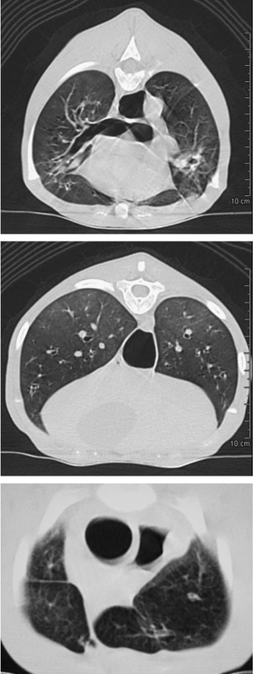
Fig. 2. (A, B) Transverse thoracic HRCT images at T5 and the T10–11 disk space, respectively. (A) Bronchial wall thickening, bronchiectasis and the abnormal visualization of bronchi or bronchioles into the periphery of the lung. (B) Diffuse bronchial wall thickening and the patchy diffuse homo- geneous opacity present in the dorsal aspect of the lungs. (C) Transverse thoracic spiral CT image at the C7–T1 disk space. Note the rounded ap- pearance of the cranial lung lobe margins. HRCT, high-resolution computed tomography; CT, computed tomography.
How did we do? |
Disclaimer: The summary generated in this email was created by an AI large language model. Therefore errors may occur. Reading the article is the best way to understand the scholarly work. The figure presented here remains the property of the publisher or author and subject to the applicable copyright agreement. It is reproduced here as an educational work. If you have any questions or concerns about the work presented here, reply to this email.