- Veterinary View Box
- Posts
- Just a classic....
Just a classic....
Journal of the American Veterinary Medical Association (JAVMA) 2016
Robert M. Kirberger
Background
Diskospondylitis is an infection of the intervertebral disk and adjacent vertebral endplates that can be caused by bacterial or fungal agents. It is typically reported in adult male, large-breed dogs, with clinical signs including spinal hyperesthesia, paresis, fever, and weight loss. However, reports of diskospondylitis in juvenile dogs (<6 months old) are rare, and its early imaging characteristics remain poorly described. This study aimed to identify early radiographic, ultrasonographic, and computed tomography (CT) findings in juvenile dogs with presumed diskospondylitis.
Methods
Study Design: Retrospective case series.
Study Population:
-10 client-owned juvenile dogs (<6 months old) diagnosed with presumed diskospondylitis between 2008 and 2014.
-Breeds included Labrador Retrievers (n = 3), Bull Terriers (n = 2), and single cases of American Pit Bull Terrier, Basset Hound, Border Collie, Jack Russell Terrier, and Boerboel-crossbred dogs.
Diagnostic Imaging:
-Radiographs (all 10 dogs): Evaluated for disk space narrowing, vertebral endplate lysis, and subluxation.
-Ultrasound (5 dogs): Assessed for disk swelling, loss of normal reverberation artifact, and adjacent tissue changes.
-CT (4 dogs): Used for volume rendering and multiplanar reconstructions.
Follow-Up:
Serial imaging was performed over a median of 6 weeks (range: 2–16 weeks).
Results
Clinical Presentation:
-All dogs showed vertebral pain within 3 weeks of recovering from trauma, bite wounds, or systemic illness (e.g., parvovirus, babesiosis).
-Fever was not consistently present, and leukocytosis was observed in 5 of 6 tested dogs.
Radiographic Findings:
-The earliest change was intervertebral disk space narrowing (detected in 28 disk spaces ≤2 weeks after symptom onset).
-Vertebral endplate lysis was not present initially but appeared in later stages.
-Subluxation of adjacent vertebrae was noted in 8 of 28 affected disks.
Ultrasonographic Findings:
-Hypoechoic, ventrally bulging material was seen at the affected disk site in 4/5 dogs.
-Loss of normal reverberation artifact was detected in 2 dogs before radiographic changes were evident.
CT Findings:
-Hypoattenuating (softened) vertebral endplates and altered disk coloration were identified in 3 of 4 dogs.
-In one dog, CT changes were seen before radiographic abnormalities appeared.
Limitations
Small sample size (n = 10) and retrospective study design.
Lack of culture confirmation in most cases (only 2/7 culture-positive samples).
Histopathologic confirmation was not performed to verify the inflammatory process.
Conclusions
Juvenile diskospondylitis differs from adult cases by presenting early with disk space narrowing and subluxation before endplate lysis develops. Ultrasound and CT may detect changes earlier than radiography, highlighting their diagnostic value in young dogs with spinal pain. Given the atypical imaging presentation in juveniles, diskospondylitis should be considered in young dogs with recent trauma or systemic illness, even if radiographs appear normal initially.

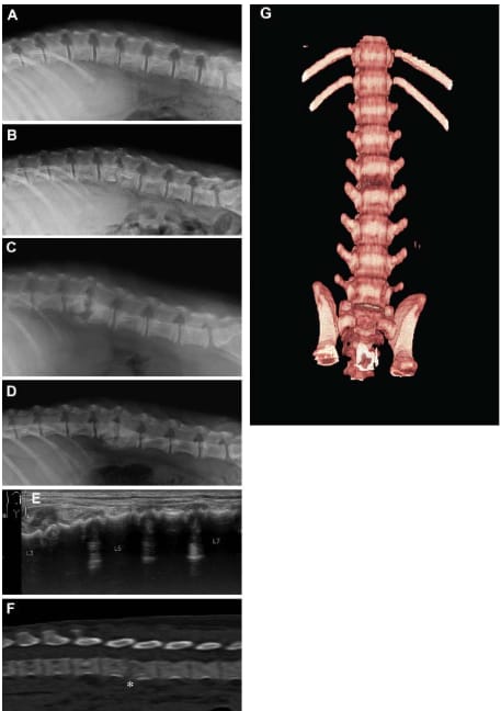
Lateral radiographic (A–D), ultrasonographic (E), and CT (F and G) images of the vertebral column in a juvenile Border Collie with presumed diskospondylitis. The patient was hospitalized for treatment of parvovirus infection at 8 weeks of age; the initial images were obtained when clinical signs suggestive of diskospondylitis developed at 9 weeks of age (A, E, F, and G), and follow-up images were obtained 1 week (B), 5 weeks (C), and 8 weeks (D) later. A—The L3–4 intervertebral disk space is narrowed with mild subluxation in the 9-week-old patient. B—At follow-up 1 week after the image in panel A was obtained, advanced disk space collapse and subluxation at L3–4 are evident. C—Five weeks after initial evaluation, changes more typical for diskospondylitis, including vertebral epiphyseal and metaphyseal lysis, grade 1 spondylosis, and vertebral malalignment are evident in the affected region. D—The 8-week follow-up image reveals filling of lytic areas with new bone and spondylosis progression to grade 3. E—A composite transabdominal ultrasonogram of the ventral aspects of L3 through L7 obtained at the same visit as the image in panel A shows ventrally bulging hypoechoic tissue at the L3–4 disk space and loss of intervertebral disk artifactual reverberation shadows as seen further caudally. F—A sagittal reconstructed lumbar CT image obtained at the initial evaluation (viewed in a bone window) reveals a poorly defined, slightly hypoattenuating L3–4 disk space (asterisk) and corresponding collapse in the interarcuate space. G—In a volume-rendered CT image of the ventral aspects of the lumbar vertebrae obtained at the initial evaluation, notice the marked change in color indicative of a region of inflammation or erosion at the L3–4 disk space.
How did we do? |
Disclaimer: The summary generated in this email was created by an AI large language model. Therefore errors may occur. Reading the article is the best way to understand the scholarly work. The figure presented here remains the property of the publisher or author and subject to the applicable copyright agreement. It is reproduced here as an educational work. If you have any questions or concerns about the work presented here, reply to this email.