- Veterinary View Box
- Posts
- Just a cool case
Just a cool case
Animals, 2025.
Miki Hirose, Lina Hamabe, Kazumi Shimada, Aki Takeuchi, Kazuyuki Terai, Aimi Yokoi, Ahmed Farag, Akari Hatanaka, Rio Hayashi, Katsuhiro Matsuura, Ryou Tanaka.
Background
Congenital left ventricular diverticulum (CLVD) is a rare congenital heart defect characterized by an outpouching of the ventricular wall, lined by endocardium and containing myocardial tissue. CLVD is often asymptomatic but may lead to complications such as arrhythmias, thrombus formation, or rupture. Due to its rarity, CLVD can be misdiagnosed as a ventricular aneurysm or pseudoaneurysm. Electrocardiosynchronous computed tomography (ECG-synchronized CT) is an advanced imaging technique that reduces motion artifacts and provides detailed cardiac imaging. This case report describes the use of ECG-synchronized CT to diagnose CLVD in a dog.
Methods
A 2-month-old female Shiba Inu presented with reduced activity and was diagnosed with pulmonary hypertension secondary to a ventricular septal defect (VSD). During follow-up echocardiography, an incidental left ventricular diverticulum was detected at the apex. To confirm the diagnosis and assess the diverticulum’s morphology, ECG-synchronized CT was performed. The study evaluated cardiac function using echocardiography, 2D speckle-tracking echocardiography (2D-STE), and Doppler analysis to characterize myocardial strain and blood flow patterns.
Results
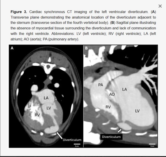
Echocardiography confirmed the presence of a 10.2 × 7.6 mm diverticulum with a 3.6 mm opening to the left ventricle. Doppler analysis revealed bidirectional blood flow into and out of the diverticulum, with peak systolic velocities of 419.1 cm/s. ECG-synchronized CT confirmed the diverticulum’s attachment to the left ventricular apex without myocardial thinning or signs of aneurysmal degeneration. 3D reconstruction images showed a thin-walled diverticulum adjacent to the sternum, with no thrombi or calcifications. Strain imaging (2D-STE) demonstrated asynchronous movement of the diverticulum but preserved global left ventricular function (global longitudinal strain = -18.7%).
Limitations
This case report is limited to a single patient, and histopathologic confirmation of the diverticulum was not available. The long-term clinical implications of CLVD remain unclear due to a lack of follow-up data. Additionally, while CT provided detailed anatomical insights, its diagnostic specificity compared to cardiac MRI remains to be established.
Conclusions
This case represents the first veterinary report of CLVD evaluated using ECG-synchronized CT. The findings highlight the potential of advanced imaging modalities in diagnosing congenital cardiac anomalies. While the diverticulum did not appear to impact cardiac function, continued monitoring is warranted due to the risk of future complications. These results support the use of ECG-synchronized CT as a valuable tool for evaluating complex cardiac anomalies in veterinary medicine.

How did we do? |
Disclaimer: The summary generated in this email was created by an AI large language model. Therefore errors may occur. Reading the article is the best way to understand the scholarly work. The figure presented here remains the property of the publisher or author and subject to the applicable copyright agreement. It is reproduced here as an educational work. If you have any questions or concerns about the work presented here, reply to this email.