- Veterinary View Box
- Posts
- MRI and Histology Link Bromethalin Toxicity to Spinal and Brain White Matter Damage in Cats
MRI and Histology Link Bromethalin Toxicity to Spinal and Brain White Matter Damage in Cats
Journal of the American Animal Hospital Association 2019
Obi Veterinary Education
February 04, 2026
Marc Kent, Eric N. Glass, Lindsay Boozer, Rachel B. Song, Elyshia J. Hankin, Renee M. Barber, Simon R. Platt, Alexander de Lahunta, Andrew D. Miller
Background:
Bromethalin is a non-anticoagulant rodenticide that causes uncoupling of oxidative phosphorylation, leading to cytotoxic and intramyelinic edema. Diagnosing bromethalin intoxication antemortem is challenging, and MRI findings associated with this toxicity have been poorly described. This report correlates MRI findings with histopathological lesions in two cats that succumbed to bromethalin intoxication.
Methods:
Two indoor-outdoor domestic shorthair cats with progressive multifocal neurologic signs underwent advanced MRI imaging, including diffusion-weighted imaging (DWI) and apparent diffusion coefficient (ADC) mapping. MRI findings were compared to histopathology of the brain and spinal cord following euthanasia. Frozen liver samples were analyzed for desmethylbromethalin, the active metabolite of bromethalin, to confirm intoxication.
Results:
MRI revealed widespread, bilateral T2 hyperintensity involving cerebral white matter, corticospinal tracts, cerebellar white matter, and spinal cord funiculi. DWI and ADC maps indicated restricted water diffusion, consistent with cytotoxic or intramyelinic edema. Histopathology demonstrated diffuse spongy degeneration (vacuolation) of white matter without axonal loss or significant gliosis, mirroring MRI changes. Trace amounts of desmethylbromethalin were detected in liver tissue, confirming exposure. Neither cat had a witnessed ingestion event; diagnosis was based on imaging suspicion and postmortem confirmation.
Limitations:
Only two cases were studied, limiting generalizability. Desmethylbromethalin levels were trace and below quantification limits, although consistent with intoxication. MRI findings were correlated only postmortem, with no antemortem serial imaging or long-term monitoring. No direct comparison to other leukoencephalopathies was made.
Conclusions:
MRI, specifically DWI and ADC sequences, is valuable for antemortem identification of cytotoxic or intramyelinic edema in suspected bromethalin intoxication. Widespread T2 hyperintensity and restricted diffusion should raise suspicion for toxic leukoencephalopathy. Early imaging could aid diagnosis and guide supportive therapy, though prevention remains critical.
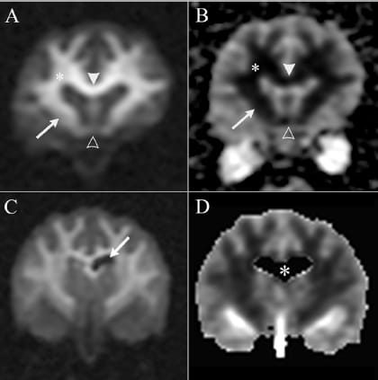
ransverse diffusionweighted
image (A, C) along with
corresponding ADC (apparent diffusion
coefficient) maps (B, D) at the
level of the rostral cerebrum of cat 1
(A, B) and the midcerebrum of cat
2 (C, D), respectively, illustrating
changes in the white matter consistent
with cytotoxic or intramyelinic
edema. (A, B) Areas of white matter,
corpus callosum (white arrowhead),
centrum semiovale (asterisk), internal
capsule (arrow), and the rostral
commissure (open arrowhead), are
hyperintense on DWI (diffusionweighted
imaging) and hypointense
on ADC maps consistent with restricted
diffusion of water. (C, D) At
the level of the midcerebrum, the
same changes are observed in the
white matter of cat 2. On the DWI
images in panel C, the normal appearance
of the cerebrospinal fluid in
the lateral ventricles is hypointense
(arrow). The cerebrospinal fluid is
conspicuous in the left lateral ventricle
as a result of slight asymmetry in
the size of the lateral ventricle. On the ADC map in panel D, the lateral ventricle and some of the surrounding parenchyma have been cropped from
the image during postprocessing (white asterisk). Normally, cerebrospinal fluid is hyperintense on ADC maps. ADC, apparent diffusion coefficient;
DWI, diffusion-weighted imaging
How did we do? |
Disclaimer: The summary generated in this email was created by an AI large language model. Therefore errors may occur. Reading the article is the best way to understand the scholarly work. The figure presented here remains the property of the publisher or author and subject to the applicable copyright agreement. It is reproduced here as an educational work. If you have any questions or concerns about the work presented here, reply to this email.