- Veterinary View Box
- Posts
- Nearly 40% of Dogs Show This CT Finding—Why Orbital Ligament Mineralization Is Likely Benign**
Nearly 40% of Dogs Show This CT Finding—Why Orbital Ligament Mineralization Is Likely Benign**
Animals 2025
Obi Veterinary Education
February 28, 2026
Ying-Ying Lo; Amélie Montenon; Aurélien Jeandel; Anne-Sophie Bedu
Background
Mineralization of the orbital ligament (OL) is occasionally observed on canine head computed tomography (CT) examinations, typically without associated clinical signs. Aside from a brief mention in the veterinary literature, its prevalence, imaging characteristics, and clinical significance have not been systematically evaluated. The authors aimed to determine the prevalence and CT features of OL mineralization in dogs and to assess associations with signalment, medical history, and concurrent mineralization. They hypothesized that OL mineralization represents a benign, incidental finding.
Methods
This retrospective descriptive study reviewed 402 canine head CT examinations performed at a single private referral hospital between January and December 2024. CT scans were evaluated for the presence of OL mineralization and characterized by laterality, symmetry, location within the ligament, size, attenuation, morphology, texture, margins, and number of lesions. Signalment (age, breed, sex, neuter status, weight), skull conformation, medical history, biochemical data (including calcium levels), CT indication, and concurrent mineralization at other sites were recorded. Statistical analyses included Chi-square or Fisher’s exact tests for categorical variables and Student’s t-test or Mann–Whitney U test for continuous variables, with Bonferroni correction applied.
Results
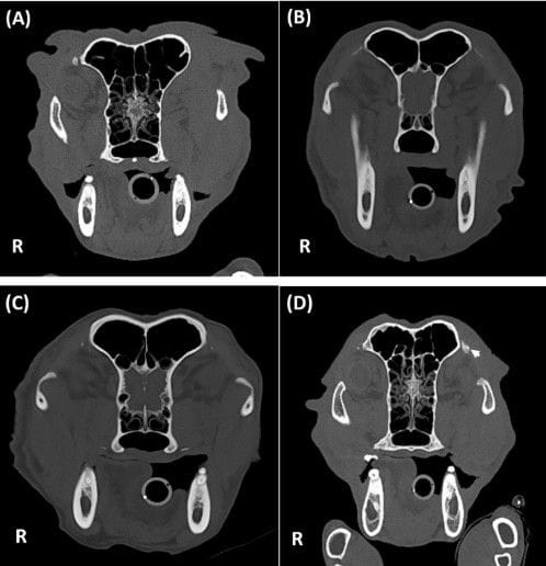
Orbital ligament mineralization was identified in 157 of 402 dogs (39.1%). Lesions were consistently dorsally located (100%) and most commonly bilateral (86.6%) and symmetrical. Morphologically, mineralization was most frequently triangular, well-defined, heterogeneous, and focal. Median lesion size was 2.48 mm and median attenuation was 793 HU. OL mineralization was significantly associated with increasing age and body weight, non-brachycephalic skull conformation, normal (non-atrophied) frontal sinuses, and concurrent mineralization at other sites, particularly in the lungs and ears. Dogs with OL mineralization were significantly older and heavier than unaffected dogs. No significant associations were found with serum calcium levels, endocrine disorders, prior corticosteroid use, chronic kidney disease, facial trauma, orbital disease, or other pathological conditions.
Limitations
The retrospective design precluded histopathological confirmation of the mineralized tissue. Image assessment relied primarily on a single observer without inter-observer reproducibility analysis. The absence of follow-up CT studies limited evaluation of lesion progression. Additionally, the relatively small number of brachycephalic dogs with OL mineralization limited subgroup comparisons.
Conclusions
Orbital ligament mineralization is a common finding in canine head CT studies, with a prevalence of 39.1% in this cohort. It is consistently dorsally located and typically bilateral and symmetrical. Its significant association with age, body weight, and other mineralization sites—without correlation to pathological conditions—supports its interpretation as a benign, incidental, age-related change without clinical significance.
How did we do? |
Disclaimer: The summary generated in this email was created by an AI large language model. Therefore errors may occur. Reading the article is the best way to understand the scholarly work. The figure presented here remains the property of the publisher or author and subject to the applicable copyright agreement. It is reproduced here as an educational work. If you have any questions or concerns about the work presented here, reply to this email.