- Veterinary View Box
- Posts
- 🐾 New CT Clues: Differentiating Inflammatory vs Neoplastic Laryngeal Masses in Dogs
🐾 New CT Clues: Differentiating Inflammatory vs Neoplastic Laryngeal Masses in Dogs
Frontiers in Veterinary Science 2025
Anna Slusarek, Annick Hamaide, Michaël Lefebvre, Marianne Heimann, Frédéric Billen, Géraldine Bolen
Background
Laryngeal tumors in dogs are rare, with squamous cell carcinoma being the most common. Clinical signs include dysphonia, stridor, cough, and dyspnea. Traditional diagnostic tools such as laryngoscopy and radiographs cannot fully characterize mass volume or margins. Computed tomography (CT) is increasingly used for diagnosis and surgical planning, but few studies directly compare the CT features of inflammatory versus neoplastic laryngeal lesions. This study aimed to describe these features and test the hypothesis that CT characteristics and lymph node enlargement could help differentiate between the two conditions.
Methods
This was a retrospective, multicenter case series including dogs with laryngeal masses diagnosed via cytology or histopathology and evaluated with CT between 2013 and 2024. Exclusion criteria included masses not originating from the larynx. CT images were reviewed by a board-certified radiologist and a resident, blinded to diagnosis. Lesions were assessed for shape, margins, growth pattern, cavitation, mineralization, cartilage invasion, and lymph node involvement. Measurements were performed on post-contrast images.
Results
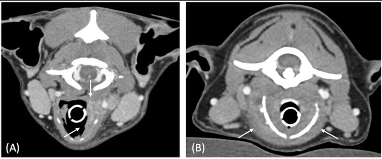
Seven dogs were included (median age 9 years). Four cases were neoplastic (all carcinomas) and three inflammatory. Dysphonia was the most common clinical sign. CT findings revealed that all neoplastic lesions appeared ovoid-shaped, whereas inflammatory lesions presented as laryngeal thickening. Both lesion types shared features such as ill-defined margins, heterogeneous contrast enhancement, cavitation (2/4 neoplastic, 2/3 inflammatory), and occasional mineralization. Cartilage destruction was observed in both groups. Regional lymphadenopathy was present in all but one case, with heterogeneous enhancement in tumor cases and more homogeneous enhancement in inflammatory cases.
Limitations
The main limitation was the small sample size, due to the rarity of laryngeal masses and the fact that CT is not a first-line diagnostic tool. The retrospective design and use of different CT machines introduced variability, though this did not affect mass shape. Some clinical data were incomplete, such as lymph node sampling results.
Conclusions
This study is the first to compare canine inflammatory and neoplastic laryngeal lesions using CT. The shape of the mass was the most reliable differentiating factor: diffuse laryngeal thickening suggested inflammation, while ovoid lesions suggested neoplasia. Other CT features overlapped between groups. Importantly, lymphadenopathy did not reliably distinguish the two etiologies. CT may therefore support differential diagnosis in dogs with laryngeal masses and guide further diagnostic and management decisions.

Figure 2. Postcontrast transverse CT images of case 3 (A) and case 6 (B), illustrating the thickened aspect of the laryngeal lesions (white arrows). Both cases had laryngeal masses with mixed external and internal growth pattern, characterized by an increase in thickness on each side of the cartilage. Window width = 400 HU, window level = 40 HU.
How did we do? |
Disclaimer: The summary generated in this email was created by an AI large language model. Therefore errors may occur. Reading the article is the best way to understand the scholarly work. The figure presented here remains the property of the publisher or author and subject to the applicable copyright agreement. It is reproduced here as an educational work. If you have any questions or concerns about the work presented here, reply to this email.