- Veterinary View Box
- Posts
- New MRI Study Links Utricle Asymmetry to Vestibular Disease in Senior Dogs
New MRI Study Links Utricle Asymmetry to Vestibular Disease in Senior Dogs
VRU 2025
Sungjun Won, Junghee Yoon
Background
Idiopathic vestibular syndrome (IVS), also known as “old dog vestibular disease,” is a common cause of acute unilateral peripheral vestibular dysfunction in older dogs. Characterized by head tilt, ataxia, and nystagmus, IVS typically resolves without treatment within weeks. However, its pathogenesis remains unclear. The study aimed to evaluate whether morphological asymmetry of the utricle—a balance-sensing structure of the inner ear—can be detected by MRI in dogs affected by IVS, suggesting potential vestibular system atrophy.
Methods
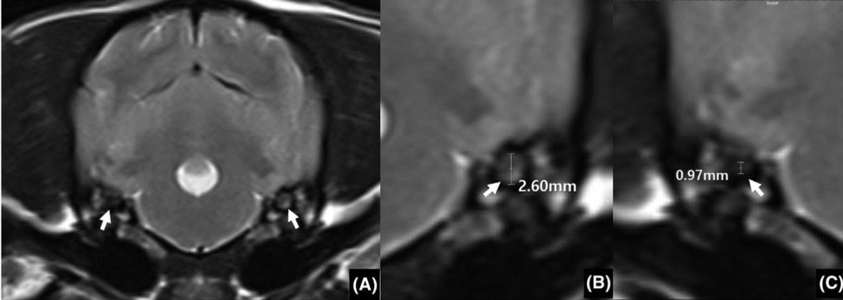
This retrospective cross-sectional study analyzed MRI scans of 137 dogs diagnosed with IVS between 2016 and 2019. After excluding other vestibular diseases and imaging artifacts, 101 dogs with IVS and 31 age-matched controls without vestibular signs were included. Transverse T2-weighted MRI images were used to measure utricle diameters bilaterally. The utricle asymmetricity ratio (UAR)—the ratio of the smaller to the larger utricle diameter—was calculated. Statistical analyses compared UARs between groups and evaluated correlations with age, sex, and symptom direction consistency.
Results
Dogs with IVS showed significantly greater utricle asymmetry than control dogs (median UAR 0.83 vs. 0.98, P < 0.01). Within the IVS group, dogs whose smaller utricle correlated with the side of clinical signs (“consistent group”) had lower UARs (0.82) compared with inconsistent cases (0.90, P < 0.01). No correlation was found between UAR and age, sex, or head-tilt direction. In dogs with UAR < 0.73, utricle size reduction always matched the side of clinical signs, suggesting meaningful structural asymmetry.
Limitations
The study was retrospective and lacked histopathologic confirmation of utricular atrophy. Technical MRI limitations (e.g., slice thickness, head obliquity) led to exclusion of 36 cases. Breed size variability prevented absolute utricle size comparisons. A more refined MRI protocol with higher spatial resolution could improve future measurements.
Conclusions
MRI revealed significant utricle diameter asymmetry in dogs with IVS compared to unaffected dogs. These findings suggest that IVS may involve structural atrophy of the peripheral vestibular system. Advanced imaging of the inner ear could enhance diagnostic accuracy and understanding of vestibular dysfunction in dogs.

Transverse T2 weighted image of the idiopathic vestibular syndrome (IVS). The oval shape of the utricle was obviouslyasymmetrical (arrow) in the IVS patient (A). Measurement of the utricle size in the largest side (B) and smallest side (C) is indicated by a whitearrow. The utricle asymmetricity ratio (UAR) was calculated by dividing the largest to the smallest utricle diameter
How did we do? |
Disclaimer: The summary generated in this email was created by an AI large language model. Therefore errors may occur. Reading the article is the best way to understand the scholarly work. The figure presented here remains the property of the publisher or author and subject to the applicable copyright agreement. It is reproduced here as an educational work. If you have any questions or concerns about the work presented here, reply to this email.