- Veterinary View Box
- Posts
- New Radiographic Angles Improve Detection of SI Luxation in Cats
New Radiographic Angles Improve Detection of SI Luxation in Cats
JSAP 2025
T. Kasa, A. Danielski, M. A. Solano, F. Santoro, V. Volckaert
Background
Sacroiliac (SI) luxation is a common traumatic injury in cats and often accompanies other pelvic fractures. Accurate diagnosis is vital for treatment planning, yet traditional radiography can yield ambiguous findings, especially in unilateral cases with subtle displacement. A quantitative method originally developed for dogs offered promise for more objective evaluation. This study aimed to validate and adapt that technique for feline patients by assessing its diagnostic accuracy and inter-observer reliability.
Methods
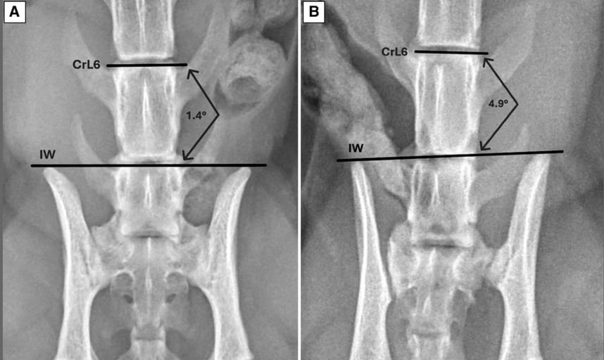
The study was conducted in two phases. Phase one involved 20 radiographs from cats with normal SI joints and 20 with confirmed unilateral SI luxation. Angles were measured between a line tangential to the cranial iliac wings and three vertebral landmarks: cranial and caudal endplates of L6, and cranial endplate of L7. In phase two, 60 radiographs (30 normal, 30 luxated) were blindly assessed twice by three observers with varying experience levels, measuring the same angles. Diagnostic performance was evaluated using ROC analysis, and intra-/inter-observer reliability was assessed using intraclass correlation coefficients.
Results
All three measured angles—CrL6, CdL6, and CrL7—were significantly greater in the luxation group. Optimal cut-off angles were identified as ≥2.3° for CrL6 and ≥2.4° for CdL6 and CrL7. These thresholds yielded high sensitivity (86–90%) and specificity (94–97%), with CrL6 demonstrating the highest predictive values. Importantly, measurement reliability was excellent across observers and time points, regardless of experience. Pelvic positioning (straight vs. oblique) did not significantly affect diagnostic performance.
Limitations
This was a retrospective, single-center study with a limited sample size. Only unilateral luxations were included, excluding more complex or bilateral cases. Although the images were cropped to reduce bias, this does not reflect clinical reality where full anatomical context is usually available. Measurement accuracy may be compromised in cases involving lumbar or iliac fractures.
Conclusions
The adapted radiographic technique offers a practical, reproducible, and accurate method for identifying unilateral SI luxation in cats. CrL6 appears most clinically useful due to anatomical clarity and excellent predictive value. These findings support broader use of quantitative angular measurements in primary care radiographic evaluation of feline pelvic trauma.

Ventrodorsal radiograph of the normal pelvis of a cat (A) and of the pelvis with unilateral sacroiliac (SI) luxation (B). The angle measured between a line parallel to the cranial endplate of the sixth lumbar vertebra (CrL6) and the line tangential to the ilium wings is 0.9° for the normal pelvis and 5.4° for the pelvis with unilateral SI luxation.
How did we do? |
Disclaimer: The summary generated in this email was created by an AI large language model. Therefore errors may occur. Reading the article is the best way to understand the scholarly work. The figure presented here remains the property of the publisher or author and subject to the applicable copyright agreement. It is reproduced here as an educational work. If you have any questions or concerns about the work presented here, reply to this email.