- Veterinary View Box
- Posts
- New Sonographic Insights into Feline Duodenal Papillae
New Sonographic Insights into Feline Duodenal Papillae
Journal of Small Animal Practice 2025
R. Leppin
Background: The anatomical and functional interplay between the common bile duct, main pancreatic duct, and duodenal papillae in cats has implications for conditions like triaditis and pancreatitis. Limited sonographic data exists on these structures. This study aimed to define the detection frequency, sonoanatomy, and reference values for the duodenal papillae and associated structures.
Methods: A prospective case-controlled study was conducted on 50 client-owned cats. High-frequency ultrasonography (15 MHz) was used to examine the major (MADP) and minor (MIDP) duodenal papillae, documenting their anatomical features, dimensions, and connections to the biliary and pancreatic ducts. Statistical analyses established normal reference ranges.
Results: The MADP was identified in 100% of cases, while the MIDP was found in 10%. The common bile duct and main pancreatic duct were consistently visualized entering the duodenum. Calculi were detected in 12% of cats, with some cases demonstrating spontaneous passage into the duodenum. The MIDP and its associated accessory pancreatic duct were observed to provide an alternative outflow pathway, potentially mitigating pancreatic duct overpressure.
Limitations: The study lacked histopathological validation, and reference values were based only on clinically healthy cats. The detection rate of the MIDP may be underestimated due to sonographic limitations.
Conclusions: High-resolution ultrasonography effectively visualizes the feline duodenal papillae, offering diagnostic and therapeutic implications for conditions like cholestasis and pancreatitis. The MIDP may play a protective role in pancreatic health, warranting further investigation

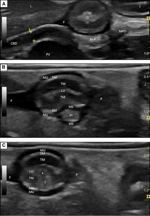
Transverse ultrasonographic images of the MADP and connected structures obtained from a 12-year-old European shorthair cat. Dorsal is to the bottom and cranial to the left of the images. (A) The CBD and the MPD appear each as a tubular structure with parallel echogenic walls and anechoic content. The area (B + P) dorsal to the duodenum can be seen, in which both ducts turn at a right angle to the right lateral aspect of the cat and run together in a parallel arrangement out of the plane of the image. (B) The PI can be seen. (C) The PS (◊) and the MADP (▪) can be seen. D Duodenum, F Free fluid, J Jejunum, L Liver, LU Lumen of the duodenum, MS Muscle sheath, MU Muscularis, P Body of the pancreas, SM Submucosa, PV Portal vein, TM Mucosa.
How did we do? |
Disclaimer: The summary generated in this email was created by an AI large language model. Therefore errors may occur. Reading the article is the best way to understand the scholarly work. The figure presented here remains the property of the publisher or author and subject to the applicable copyright agreement. It is reproduced here as an educational work. If you have any questions or concerns about the work presented here, reply to this email.