- Veterinary View Box
- Posts
- New Study: Hepatolithiasis in Dogs After Cholecystectomy — Benign and Low-Risk Findings
New Study: Hepatolithiasis in Dogs After Cholecystectomy — Benign and Low-Risk Findings
J Am Vet Med Assoc. 2025
Emmy Y. Luo, Ian Porter, Meredith Miller, Nicole Buote
Background
Hepatolithiasis, or intrahepatic bile duct calculi, is an uncommon condition in dogs often discovered incidentally during diagnostic imaging. Although associated with significant complications in humans, such as cholangiocarcinoma and recurrent cholangitis, its clinical importance in dogs remains unclear. Prior studies indicated that hepatolithiasis in canines might not cause hepatobiliary dysfunction, yet the long-term outcomes after cholecystectomy have not been evaluated. This study aimed to assess the long-term clinical significance of hepatolithiasis identified in dogs undergoing cholecystectomy.
Methods
This retrospective case series reviewed dogs treated at the Cornell University College of Veterinary Medicine between January 2014 and May 2024. Dogs were included if they underwent cholecystectomy and had evidence of hepatolithiasis or intrahepatic mineralization on preoperative ultrasound or CT. Medical records were analyzed for clinical presentation, diagnostic findings, surgical details, histopathology, complications, and follow-up outcomes. Long-term follow-up was obtained via medical records and owner telephone interviews to assess postoperative biliary disease or obstruction.
Results
Of 183 dogs that underwent cholecystectomy, 14 (8%) were diagnosed with hepatolithiasis preoperatively. The median age was 11 years, and the median weight was 7.3 kg. Most were small-breed dogs presenting with anorexia or vomiting. Liver biopsies showed hepatitis or cholangiohepatitis in 64% of cases. None of the hepatoliths were surgically removed. Two dogs (14%) died perioperatively, but none of the 12 survivors developed biliary obstruction or hepatolith migration during a median follow-up of 538 days (range, 147–3,316). Long-term medications were minimal, and no dog required secondary surgery. Follow-up imaging, when performed, showed no progression or movement of hepatoliths.
Limitations
The small sample size and retrospective design limited statistical power and generalizability. Incomplete medical records and reliance on owner recall introduced potential bias. Only a subset of dogs received follow-up imaging, restricting evaluation of subclinical progression. Being a single-institution study, regional variations could not be assessed.
Conclusions
Hepatolithiasis in dogs undergoing cholecystectomy did not result in postoperative biliary obstruction or adverse outcomes during the follow-up period. The condition appears clinically insignificant in this context and should not alter surgical decision-making. Routine hepatolith removal is unnecessary unless clinically warranted. Larger, multi-institutional studies with extended follow-up are recommended to confirm these findings.

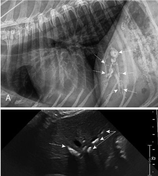
Radiographic and ultrasound images of a dog with identified hepatoliths that underwent a cholecystectomy between January 2014 to May 2024. A—A right lateral radiograph showing moderate hepatoliths (arrows). B—An ultrasound image of the same dog with the mineralizations visualized as linear hyperechoic regions (arrows) within the liver that cast an acoustic shadow.
How did we do? |
Disclaimer: The summary generated in this email was created by an AI large language model. Therefore errors may occur. Reading the article is the best way to understand the scholarly work. The figure presented here remains the property of the publisher or author and subject to the applicable copyright agreement. It is reproduced here as an educational work. If you have any questions or concerns about the work presented here, reply to this email.