- Veterinary View Box
- Posts
- New Study Reveals Surgical Fix for Rare Guinea Pig Dental Disorder
New Study Reveals Surgical Fix for Rare Guinea Pig Dental Disorder
Veterinary Clinics of North America: Exotic Animal Practice 2025
Obi Veterinary Education
October 03, 2025
Justyna Ignaszak-Dziech, DVM; Vladimir Jekl, DVM, PhD, DECZM
Background
Macrodontia is a rare dental anomaly characterized by the enlargement and abnormal structural development of one or more teeth. While well-documented in humans and some domestic animals, its occurrence in guinea pigs is underreported and typically mislabeled. Given guinea pigs' unique continuously growing (elodont) dentition, recognition and classification of such anomalies have proven challenging. This study aimed to review and systematize current knowledge on macrodontia in guinea pigs, examining histological characteristics, diagnostic strategies, and treatment approaches.
Methods
The study involved a review of literature and clinical case studies, including histological examinations and imaging (radiography and CT) of affected teeth. It also incorporated clinical data from veterinary examinations, diagnostic imaging procedures (especially cone-beam computed tomography), and surgical interventions including apicoectomy. Histologic features were compared to odontomas and other odontogenic anomalies.
Results
Macrodont teeth showed disorganized arrangements of typical dental tissues, including osteodentin and cementum, with deformities localized primarily in the apex and crown. These changes mimicked compound or complex odontomas but were confined to the pulp cavity and did not extend beyond the alveolus. Most commonly affected were the M2 and M3 molars, particularly in the maxilla. Clinical signs included malocclusion, facial swelling, and occlusal surface defects. Radiographic findings revealed crown enlargement, loss of enamel contrast, and widened periodontal spaces. CT imaging offered superior detail, with 85% of cases exhibiting significant alveolar deformation.
Limitations
The etiology of macrodontia remains undetermined. Although parallels with trauma, vitamin deficiencies, and inflammation have been suggested based on data from other species, no causal relationship has been confirmed in guinea pigs. Additionally, histological comparisons are limited by the lack of standardized veterinary dental classification systems.
Conclusions
Macrodontia in guinea pigs is a significant dental pathology contributing to malocclusion and secondary complications. Accurate diagnosis requires a combination of clinical examination, endoscopy, and advanced imaging. CT is particularly useful for surgical planning. Apicoectomy is preferred over extraction to minimize complications and promote safe alveolar healing. Continued research is needed to clarify etiology and refine treatment protocols.
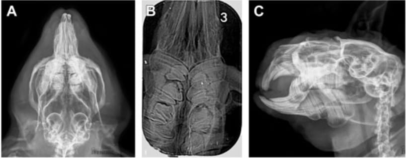
Radiography of a guinea pig with macrodont teeth (A–C). (A) Isolated view of a maxilla according to Bohmer.8 A homogeneous structural alteration is visible in the mesial part of the right maxillary M2, leading to an enlarged crown outline. The mesial periodontal space is widened secondary to crown substance loss. The left maxillary M2 also displays features of macrodontia. Crown margins are uneven, pulp cavities are not visible, and both mesial and distal periodontal spaces are significantly widened. The left maxillary M3 is clearly displaced caudally and rotated due to the enlarged M2 crown outline. (B) Dorsoventral intraoral projection of the maxilla. Homogeneous structural changes are visible in the mesial parts of the right maxillary M1 and M2. Crown outlines are enlarged, with uneven mesial edges and a minor defect contributing to periodontal space widening. Similar changes are seen in the mesial parts of left M2 and M3. Loss of dental substance in the mesial portion of M2 leads to periodontal space widening. (C) Latero-oblique projection of the right mandible. Marked crown enlargement of M2 with blurred structural boundaries. The mesial pulp cavity shows increased radiodensity. Periodontal spaces are widened, and a longitudinal radiolucent line running through the crown suggests a possible fracture. Periapical features of periosteal reaction are visible. The M1 and M3 crowns show secondary rotation, giving the impression of reduced tooth size.
How did we do? |
Disclaimer: The summary generated in this email was created by an AI large language model. Therefore errors may occur. Reading the article is the best way to understand the scholarly work. The figure presented here remains the property of the publisher or author and subject to the applicable copyright agreement. It is reproduced here as an educational work. If you have any questions or concerns about the work presented here, reply to this email.