- Veterinary View Box
- Posts
- Poor man CT brings no benefits....
Poor man CT brings no benefits....
Veterinary Radiology & Ultrasound, 2017
Danielle Butler, Sarah Nemanic, Jennifer J. Warnock
Background
Fractures of the canine tarsus can be challenging to diagnose due to the complex anatomy and overlapping structures on radiographs. Accurate detection is essential for selecting appropriate treatment, as undiagnosed fractures can lead to osteoarthritis and chronic lameness. This study aimed to compare the accuracy, sensitivity, and specificity of computed tomography (CT) and radiography (ten-view and two-view studies) in detecting traumatic tarsal fractures in dogs.
Methods
A prospective experimental study was conducted using 10 cadaveric canine hind limbs from medium to large breed dogs. Each limb underwent CT scanning and ten-view radiographic imaging before and after fractures were induced using a hydraulic press. Two independent observers evaluated all imaging modalities for fracture detection, with post-dissection findings serving as the gold standard. The two-view radiographic studies (dorsoplantar and lateromedial views) were retrospectively extracted from the ten-view studies and analyzed separately.
Results
All limbs sustained fractures, with the talus and calcaneus being the most commonly affected bones (n=7 each). CT demonstrated higher sensitivity (77%) compared to ten-view radiographs (57%), though specificity was similar (97% vs. 98%). No significant improvement in fracture detection was observed between ten-view and two-view radiographic studies (57% vs. 55% sensitivity, both 98% specificity). CT was particularly advantageous for detecting fractures of the fourth tarsal and central tarsal bones. Post-dissection image review revealed that some fractures initially missed on CT were due to observer error rather than modality limitations.
Limitations
This study used cadaveric limbs, which lack soft tissue swelling and post-traumatic changes that could affect clinical imaging interpretation. The fractures were induced under controlled conditions, which may not fully replicate those seen in live patients. Additionally, a small sample size limits generalizability.
Conclusions
CT is superior to radiography for detecting fractures of the canine tarsus, particularly for subtle or complex fractures. However, two-view radiographic studies performed similarly to ten-view studies, suggesting that extensive radiographic series may not provide additional diagnostic benefit. These findings support the use of CT in cases where fractures are suspected but not clearly identified on radiographs.

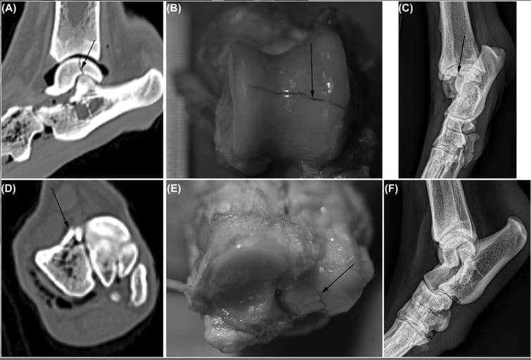
Postfracture images of Limb 1. Based on dissection, this limb sustained a fracture of the trochlear ridges of the talus (B, arrow) as well as nondisplaced chip fracture of the 4th tarsal bone (E, arrow). The fracture of the talus was seen on sagittal plane computed tomography (CT) images (A, arrow) as well as multiple radiographic projections (C, F, arrows). The nondisplaced fracture of the 4th tarsal bone was seen on transverse CT images (D, arrow), but not on radiographs (C, F). Bone algorithm CT images are displayed in a bone window and level (W: 2000, L: 400). Of the ten-view radiographic study, the dorsomedial-plantarolateral (C), and lateromedial (F) projections are shown
How did we do? |
Disclaimer: The summary generated in this email was created by an AI large language model. Therefore errors may occur. Reading the article is the best way to understand the scholarly work. The figure presented here remains the property of the publisher or author and subject to the applicable copyright agreement. It is reproduced here as an educational work. If you have any questions or concerns about the work presented here, reply to this email.