- Veterinary View Box
- Posts
- Radiographic Clues in Equine Carpal Lameness—What’s Incidental vs. Pathologic?
Radiographic Clues in Equine Carpal Lameness—What’s Incidental vs. Pathologic?
VRU 2010
Valerie Simon, Sue J. Dyson
Background:
Radiologic interpretation of the equine carpus is challenging due to numerous anatomic variations, such as the presence of supernumerary carpal bones and various shapes of the ulnar carpal bone. This study aimed to assess the prevalence and clinical significance of radiologic variants in horses with carpal lameness and controls, focusing on potential correlations with breed, gender, discipline, and lameness origin.
Methods:
A total of 286 sets of carpal radiographs from 222 horses were reviewed. These included horses of various breeds, disciplines, and both genders. Radiographs were categorized based on the presence or absence of lameness and localized sources of pain. Variants assessed included presence of the first and fifth carpal bones, radiolucencies in various carpal and metacarpal bones, and the shape of the ulnar carpal bone. Chi-square tests and Bonferroni corrections were applied to evaluate associations.
Results:
The first carpal bone was present in 29% of limbs, more commonly in Thoroughbred-cross horses, while the fifth carpal bone was rare (1.4%). Radiolucent areas in the second carpal bone were more frequent in females but not breed-associated. The ulnar carpal bone displayed five shape variants, most commonly bilobed (71.3%). Radiolucencies and osseous opacities on the palmaromedial aspect of the ulnar carpal bone occurred in 2.4% and 1.4% of limbs, respectively, and were not associated with lameness or any demographic variables. No radiographic variants evaluated were statistically associated with carpal lameness.
Limitations:
The study population was drawn from referral centers, which may not represent the general equine population. Variability in discipline and lameness group sizes may affect statistical power. No standardized object for radiographic magnification correction was used.
Conclusions:
Most radiologic anatomic variations of the carpus, including those involving the ulnar carpal bone, are not associated with lameness and likely represent incidental findings. Awareness of these variants is important for accurate radiographic interpretation and to avoid over-diagnosis of pathology.

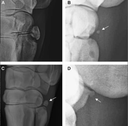
(A) Dorsomedial–palmarolateral oblique radiograph. There is a large first carpal bone articulating with the second carpal and second metacarpal bones and radiolucencies in the second carpal and second metacarpal bones. (B) Dorsomedial–palmarolateral oblique radiograph. There is a much smaller osseous opacity (first carpal bone) palmar to the second carpal bone (arrow) and radiolucencies in the second carpal and second metacarpal bones. (C) Dorsolateral–palmaromedial oblique radiograph. There is a fifth carpal bone (arrow). (D) Weight-bearing lateromedial radiograph. Note the separate center of ossification palmar to the proximal row of carpal bones (arrow).
How did we do? |
Disclaimer: The summary generated in this email was created by an AI large language model. Therefore errors may occur. Reading the article is the best way to understand the scholarly work. The figure presented here remains the property of the publisher or author and subject to the applicable copyright agreement. It is reproduced here as an educational work. If you have any questions or concerns about the work presented here, reply to this email.