- Veterinary View Box
- Posts
- Rare Cervical Spine Mass in Dog Resolved After Surgery—No Recurrence for 6 Years
Rare Cervical Spine Mass in Dog Resolved After Surgery—No Recurrence for 6 Years
Animals 2025
Obi Veterinary Education
January 24, 2026
Yoshiyuki Inoue, Rie Kitoh, Moe Satoh, Yuki Yoshigae, Kazumi Nibe, Kazuyuki Uchida, Satoru Matsunaga
Background:
Inflammatory pseudotumors (IPTs) are benign, tumor-like lesions characterized by inflammatory cell infiltration and mesenchymal proliferation. Though commonly reported in the orbits or lungs of humans and animals, spinal involvement is extremely rare. This report describes the clinical course, imaging findings, surgical management, and long-term outcome of an epidural IPT in the cervical spine of a young Bernese Mountain Dog.
Methods:
A 3-year, 9-month-old female Bernese Mountain Dog presented with acute right-sided hemiparesis. Neurologic examination localized the lesion to the C1–C5 spinal segments. MRI revealed a homogeneously enhancing extradural mass at C3–C4 compressing the spinal cord. The mass was removed via dorsal laminectomy. Intraoperative cytology was suggestive of lymphoma, but histopathology and immunohistochemistry confirmed a diagnosis of IPT. Postoperative follow-up included MRI and long-term monitoring via owner report.
Results:
The mass was completely excised with minimal adhesion to the dura. Histopathology revealed proliferation of spindle cells and infiltration by lymphocytes, plasma cells, and macrophages without significant atypia or monoclonality. Neurological signs resolved by postoperative day 12, and MRI at day 63 confirmed no recurrence. The dog remained neurologically normal at a 6-year follow-up. No adjuvant treatment was required after surgery, and the clinical outcome was excellent.
Limitations:
As a single case report, findings are not generalizable. The retrospective nature limited preoperative diagnostics, and definitive diagnosis required postoperative tissue analysis. The underlying etiology of the IPT remains unknown. Diagnostic imaging and cytology alone were insufficient to distinguish IPT from other neoplasms.
Conclusions:
This case illustrates that epidural IPT in the canine cervical spine, though rare, can present with acute neurologic deficits and mimic neoplasia on imaging. Complete surgical excision can result in rapid and sustained clinical improvement. Due to its benign nature and favorable prognosis after removal, IPT should be considered in the differential diagnosis for extradural spinal masses in dogs, particularly young Bernese Mountain Dogs.
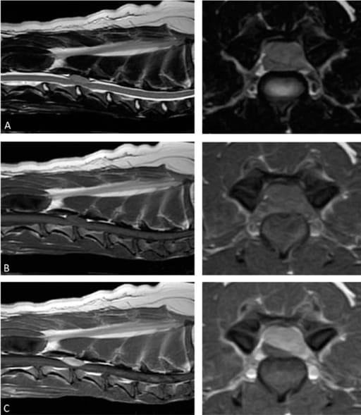
This figure is the preoperative magnetic resonance imaging of the epidural mass lesion. The mass lesion is isointense to the spinal cord parenchyma on T2-weighted images: (A) T1-weighted images; (B) the postcontrast T1-weighted images reveal a homogeneous enhancing lesion with spinal cord compression; (C) no abnormality is observed in the adjacent bone.
How did we do? |
Disclaimer: The summary generated in this email was created by an AI large language model. Therefore errors may occur. Reading the article is the best way to understand the scholarly work. The figure presented here remains the property of the publisher or author and subject to the applicable copyright agreement. It is reproduced here as an educational work. If you have any questions or concerns about the work presented here, reply to this email.