- Veterinary View Box
- Posts
- Rare Feline Biliary Disorder Uncovered: New Insights into Emphysematous Cholecystitis
Rare Feline Biliary Disorder Uncovered: New Insights into Emphysematous Cholecystitis
JSAP 2025
M. Hernandez Perello, C. Prior, M. Moloney, and C. Albuquerque
Background:
Emphysematous cholecystitis (EC) is a rare, severe gallbladder condition caused by gas-producing bacteria. While previously documented in humans and sporadically in dogs, there have been no formal case series in cats. This study aims to characterize the clinical and pathological features of EC in feline patients to inform diagnosis and treatment strategies.
Methods:
A retrospective review was conducted across 32 Diplomate-led internal medicine referral centers in the UK and Ireland. Inclusion criteria required imaging-confirmed diagnosis of EC in cats. Clinical, laboratory, imaging, cytological, histopathological, and treatment data were compiled and analyzed descriptively for five confirmed cases from 2017 to 2022.
Results:
The five cats (3 Domestic Shorthair, 2 Siamese; ages 6.5–13 years) presented with inappetence (100%), vomiting (80%), and lethargy (60%). Common lab abnormalities included leucocytosis, lymphopenia, elevated ALT, and hyperbilirubinemia. Imaging revealed gas within the gallbladder or biliary tree, gallbladder wall thickening, and choleliths. Two of five bile cultures were positive (Enterococcus spp. and Corynebacterium spp.). Histopathology indicated chronic cholecystitis and portal fibrosis without evidence of necrosis. Three cats, all of whom received medical stabilization followed by cholecystectomy, survived to discharge.
Limitations:
The study's retrospective nature, small sample size, and data heterogeneity (from different centers and labs) limit generalizability. Imaging assessments lacked centralized review. Pre-treatment with antibiotics may have led to underreporting of bacterial involvement.
Conclusions:
Feline EC is a rare but clinically significant condition that should be considered in cats with hyperbilirubinemia and elevated ALT. Diagnosis relies on imaging, and outcomes appear favorable with a combination of antibiotic therapy and surgical intervention. Further prospective studies are needed to clarify predisposing factors and optimize management protocols.

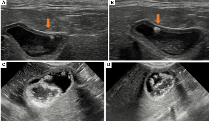
Ultrasonographic appearance of a cat diagnosed with emphysematous cholecystitis (EC) (A, B). Diffusely thickened wall of the gallbladder witha focal hyperechoic region eliciting a distal reverberation or comet tail artefact (arrow). Ultrasonographic appearance of a different cat with EC (C,D). The gallbladder lumen contains multiple small non-dependent hyperechoic foci with comet tail artefact, with a couple of echogenic foci within thewall; the gallbladder wall is diffusely thickened. Additionally, a moderate amount of heterogeneous hyper- and hypo-echoic material is present in thedependent aspect of the gallbladder.A BC D
How did we do? |
Disclaimer: The summary generated in this email was created by an AI large language model. Therefore errors may occur. Reading the article is the best way to understand the scholarly work. The figure presented here remains the property of the publisher or author and subject to the applicable copyright agreement. It is reproduced here as an educational work. If you have any questions or concerns about the work presented here, reply to this email.