- Veterinary View Box
- Posts
- Same old, same old....
Same old, same old....
Journal of Veterinary Internal Medicine 2022
Sergio A. Gomes, Mike Targett, Mark Lowrie
Discospondylitis is an infectious disease affecting the intervertebral disc space (IVDS), adjacent endplates, and vertebral bodies in dogs, causing severe spinal pain and neurological deficits. While magnetic resonance imaging (MRI) is the gold standard for diagnosis, computed tomography (CT) is increasingly available in veterinary practice. This study aimed to describe CT features of discospondylitis in dogs and evaluate how effectively these findings correlate with MRI-diagnosed discospondylitis.
Methods
Study Population:
-41 dogs diagnosed with discospondylitis via MRI at a single referral hospital (2012–2022).
-A total of 63 affected intervertebral disc sites were analyzed.
Imaging Protocol:
-MRI was used to confirm discospondylitis based on T2-weighted and contrast-enhanced findings.
-CT scans were retrospectively reviewed, with the following features evaluated:
-Endplate changes (erosion, sclerosis, osteolysis).
-Vertebral body involvement (osteolysis, periosteal reaction, spondylosis).
-IVDS narrowing, collapse, or gas accumulation (vacuum phenomenon).
-Epidural space and paraspinal soft tissue abnormalities.
Statistical Analysis:
-Descriptive analysis of CT lesion prevalence and distribution.
Results
Most common CT features of discospondylitis:
-Endplate involvement (87.3%), predominantly bilateral (94.5%).
-Endplate erosion (61.9%) and multifocal osteolysis (67.3%) were frequent.
-Periosteal proliferation (73%) and ventral spondylosis (66.7%) adjacent to affected IVDS.
-Vertebral body osteolysis (54%) involving one-third of the vertebra in most cases (85.7%).
-Abnormal IVDS appearance (42.9%), with narrowing (40.7%) or collapse (40.7%).
Less common findings:
-Local Disseminated Idiopathic Skeletal Hyperostosis (DISH) (25.4%).
-Vacuum phenomenon (23.8%) (gas within the IVDS).
-Epidural space involvement (11.1%), primarily nerve root compression at L7 (7.9%).
-Paraspinal soft tissue alterations (9.5%), but no abscess formation observed.
-Most frequently affected site: L7-S1 (25.4% of cases).
Microbial culture results:
-28.2% of cases had positive cultures, with Staphylococcus spp., Streptococcus spp., Brucella canis, and E. coli being most common.
-All dogs improved with antimicrobial therapy, confirming infectious etiology.
Limitations
Retrospective study design, with potential case selection bias.
Lack of CT specificity for early discospondylitis, as MRI remains superior for soft tissue and early bone marrow changes.
Only 28% of cases had confirmed bacterial or fungal cultures, limiting pathogen-specific conclusions.
Conclusions
CT findings of discospondylitis in dogs commonly include bilateral endplate erosion, vertebral osteolysis, and periosteal proliferation. While MRI remains the gold standard, CT can provide valuable diagnostic information, particularly when MRI is unavailable. Careful evaluation of CT scans in all three planes is essential, as some subtle cases may be missed without thorough assessment. These findings support the use of CT as a diagnostic adjunct for discospondylitis but highlight the continued need for MRI in equivocal cases.

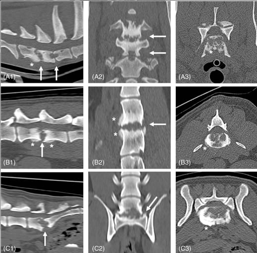
Three examples of discospondylitis in dogs on CT presenting frequently identified features: First row (A) C6-C7 and C7-T1 multiple discospondylitis (affected sites indicated by arrows), bone window: A1 sagittal, A2 dorsal, A3 transverse plane through C6-C7 disc space. At C6-C7, there is evidence of erosion of the endplates bilaterally with multifocal osteolysis, periosteal proliferation (asterisk), involvement of a third of the affected vertebral bodies with osteolysis and sclerosis; the intervertebral disc space (IVDS) was virtually enlarged. At C7-T1, there is evidence of erosion of the endplates bilaterally with focal osteolysis, and a narrow IVDS. This case had positive fungal culture for Paecilomyces spp. (possible formosus) from intrasurgical samples of both discs. Second row (B) L4-L5 discospondylitis (arrow), bone window: B1 sagittal, B2 dorsal, B3 transverse plane. There is evidence of erosion of the endplates bilaterally with multifocal osteolysis, mild periosteal proliferation (asterisk) ventral to the affected vertebral bodies without spondylosis formation, a third of the affected vertebral bodies were involved with sclerosis. The IVDS was virtually enlarged. This case had positive bacterial urine culture for E. coli. Third row (C) L7-S1 discospondylitis (arrow), bone window: C1 sagittal, C2 dorsal, C3 transverse plane. There is evidence of erosion of both endplates with multifocal coalescent osteolysis, periosteal proliferation (asterisk) without spondylosis formation, two-thirds of the affected vertebral bodies involved with osteolysis and sclerosis. The IVDS was virtually enlarged and a degree of subluxation was evident. This case had positive serological titers for Brucella canis
How did we do? |
Disclaimer: The summary generated in this email was created by an AI large language model. Therefore errors may occur. Reading the article is the best way to understand the scholarly work. The figure presented here remains the property of the publisher or author and subject to the applicable copyright agreement. It is reproduced here as an educational work. If you have any questions or concerns about the work presented here, reply to this email.