- Veterinary View Box
- Posts
- Should we MR our biliary disease dog?
Should we MR our biliary disease dog?
Veterinary Radiology & Ultrasound 2025
Reina Fujiwara, Kie Yamamoto, Masahiro Yamasaki, Koichi Ohno
Background
Diagnosing biliary and pancreatic duct diseases in dogs relies on imaging techniques such as ultrasonography, computed tomography (CT), and endoscopic retrograde cholangiopancreatography (ERCP). However, ultrasonography has operator-dependent limitations, while ERCP is invasive. In human medicine, magnetic resonance cholangiopancreatography (MRCP) has become the standard for noninvasive imaging of bile and pancreatic ducts, but data on its use in dogs is limited. This study aimed to evaluate the feasibility of MRCP for visualizing the biliary and pancreatic duct systems in healthy dogs.
Methods
Study Design: Prospective observational study.
Study Population:
-10 client-owned dogs with no history of hepatic, biliary, or pancreatic disease.
-Breeds included Chihuahuas (n = 3), Toy Poodles (n = 3), Miniature Dachshunds (n = 2), a Labrador Retriever (n = 1), and a mixed-breed dog (n = 1).
-Median age: 10 years (range: 7–14.8 years).
Imaging Protocol:
-3.0T MRI scanner (Vantage Galan 3T, Canon Medical Systems, Japan).
-Respiratory-gated, heavily T2-weighted sequences were used to visualize bile and pancreatic ducts.
-Measurements included visibility grading (Excellent, Good, Fair, Poor) and ductal diameter assessment.
Results
Detection Rates of Anatomical Structures:
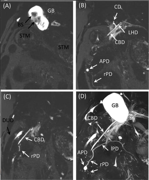
-Gallbladder: 100% detection rate (80% Excellent, 20% Good).
-Common bile duct: 80% detection rate (60% Excellent, 10% Good, 10% Fair).
-Cystic duct: 70% detection rate (40% Excellent, 20% Good, 10% Fair).
-Left and right hepatic ducts: 60% detection rate.
-Pancreatic ducts in the left and right lobes: 70% detection rate.
-Accessory pancreatic duct: 60% detection rate.
-Major pancreatic duct: 40% detection rate.
Structural Insights:
-Larger lumens were more easily visualized.
-The major pancreatic duct had the lowest visibility (40%) despite a median diameter of 1.0 mm, likely due to its secondary role in exocrine pancreatic drainage in dogs.
Limitations
-Small sample size (n = 10), limiting generalizability.
-No diseased dogs included, preventing evaluation of MRCP’s diagnostic accuracy in clinical cases.
-Single-observer image assessment, increasing potential for bias.
Conclusions
MRCP is a feasible and noninvasive method for imaging the biliary and pancreatic duct systems in dogs, with high visibility for major bile ducts but limited visualization of the major pancreatic duct. The study suggests MRCP could serve as an alternative to invasive ERCP in dogs and may be useful for preoperative planning in biliary and pancreatic diseases. Further studies should evaluate MRCP’s diagnostic accuracy in diseased patients and explore optimization techniques for improving pancreatic duct visualization.

MRCP images of Dog 7. All structures except the major pancreatic duct are excellent. A–C, Partial MIP images with a thickness of 3 mm. D, MIP image. APD, accessory pancreatic duct; BS, bile sludge; CBD, common bile duct; CD, cystic duct; DUO, duodenum; GB, gallbladder; LHD, left hepatic duct; lPD, pancreatic duct in the left lobe; MIP, maximum intensity projection; rPD, pancreatic duct in the right lobe; STM, stomach.
How did we do? |
Disclaimer: The summary generated in this email was created by an AI large language model. Therefore errors may occur. Reading the article is the best way to understand the scholarly work. The figure presented here remains the property of the publisher or author and subject to the applicable copyright agreement. It is reproduced here as an educational work. If you have any questions or concerns about the work presented here, reply to this email.