- Veterinary View Box
- Posts
- Should you CT or MRI for spinal trauma patients?
Should you CT or MRI for spinal trauma patients?
Veterinary Radiology & Ultrasound, 2019
Aitor Gallastegui, Emma Davies, Allison L. Zwingenberger, Stephanie Nykamp, Mark Rishniw, Philippa J. Johnson
Background
Accurate evaluation of vertebral fractures in canine trauma patients is essential for determining spinal stability and guiding treatment. Computed tomography (CT) is considered the gold standard for assessing osseous structures, while magnetic resonance imaging (MRI) is used for evaluating soft tissues. Some studies suggest that MRI may be sufficient for detecting vertebral fractures, potentially reducing the need for CT. This study aimed to compare the accuracy of MRI and CT in detecting vertebral fractures and to assess interobserver agreement when using MRI alone.
Methods
A multicenter retrospective study included 29 dogs with CT-confirmed vertebral fractures and 4 dogs without fractures as controls. All dogs underwent CT and MRI within 48 hours of each other. Two blinded observers evaluated 128 vertebrae for fractures using MRI, and results were compared to CT findings. Fractures were categorized as stable or unstable based on the involvement of multiple vertebral compartments. Interobserver agreement was assessed using Cohen’s kappa statistic.
Results
MRI had high sensitivity (87.9–89.2%) and specificity (79.0–82.4%) for detecting fractured vertebrae, but interobserver agreement was only moderate (κ = 0.584). MRI performance improved when detecting unstable fractures, with higher specificity (96.5–99.1%) and substantial interobserver agreement (κ = 0.650). However, complete agreement between MRI and CT for exact fracture location was low (14.3–32.6%), with up to 79% of fractures in certain vertebral structures being missed on MRI. Transverse process fractures were the most frequently overlooked.
Limitations
This retrospective study included imaging from multiple institutions with variable MRI protocols, potentially affecting diagnostic accuracy. The lack of histopathologic confirmation and the assumption that CT was the gold standard introduce potential bias. Additionally, MRI assessment of vertebral fractures remains dependent on observer expertise.
Conclusions
MRI can identify vertebral fractures in dogs with reasonable accuracy but lacks the precision of CT for determining fracture morphology and location. While MRI may be useful when CT is unavailable, it should not replace CT for assessing osseous spinal trauma. MRI is most reliable for detecting unstable fractures that require surgical intervention, but small or peripheral fractures are frequently missed.

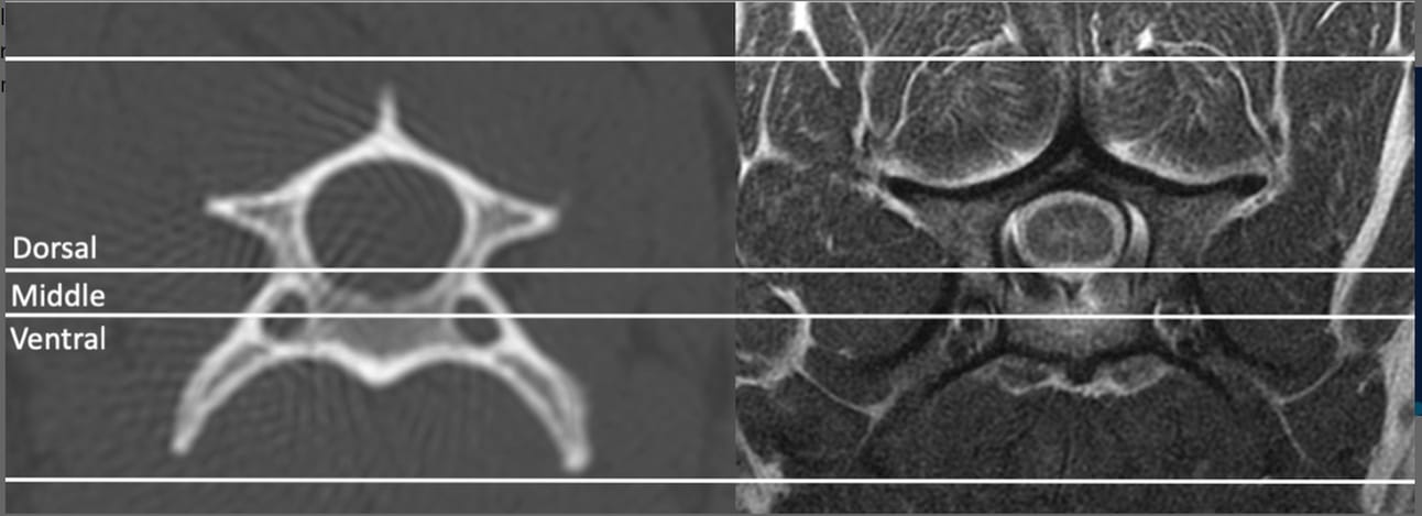
Transverse plane bone algorithm CT (0.625 mm slice thickness, window length 600, window width 2900) and corresponding transverse proton density MRI (5 mm slice thickness) images of a cervical vertebra demonstrating the division of the vertebra into three compartments. The dorsal compartment includes the lamina, dorsal pedicles, cranial and caudal articular processes, dorsal ligamentous complex, articular process joint capsule, and interarcuate, interspinous, and supraspinous ligaments. The middle compartment includes the dorsal aspect of the body, ventral pedicles, dorsal annulus, and dorsal longitudinal ligament. The ventral compartment includes the ventral body, lateral and ventral annulus, ventral longitudinal ligament and nucleus pulposus. When two or more of these compartments is fractured or ruptured then the column is considered unstable
How did we do? |
Disclaimer: The summary generated in this email was created by an AI large language model. Therefore errors may occur. Reading the article is the best way to understand the scholarly work. The figure presented here remains the property of the publisher or author and subject to the applicable copyright agreement. It is reproduced here as an educational work. If you have any questions or concerns about the work presented here, reply to this email.