- Veterinary View Box
- Posts
- Those old dog lungs
Those old dog lungs
Veterinary Radiology & Ultrasound, 2017.
Natasha L. Hornby, Christopher R. Lamb.
Background
Age-related changes in the lungs have been documented in humans and dogs, with histological studies suggesting alterations such as fibrosis, bronchial cartilage calcification, and emphysema. Radiographic studies in dogs have also described increased lung opacity and pleural thickening with age. However, no study has systematically assessed whether these changes are detectable on computed tomography (CT). This study aimed to determine if the CT appearance of the lungs differs between young and old dogs, potentially influencing diagnostic interpretation.
Methods
A retrospective case-control study analyzed thoracic CT scans from 42 young dogs (0.3–4.8 years) and 47 old dogs (9–15.1 years) without known thoracic disease. All CT scans were performed using the same multislice scanner with standardized settings. An experienced radiologist, blinded to the dogs’ ages, evaluated lung attenuation, presence of ground glass opacity, cysts, bronchial thickening or dilation, interlobular septal thickening, and lung lobe collapse. Statistical analyses assessed differences in imaging findings between young and old dogs.
Results
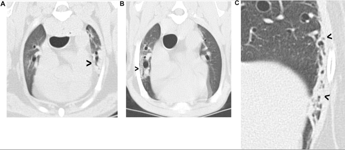
CT findings showed minimal differences between young and old dogs. The most notable age-related change was a higher prevalence of heterotopic bone in old dogs (62% vs. 14%, P < 0.001). Lung lobe collapse was significantly associated with older age, larger body weight, and anesthesia. No significant differences were found in lung attenuation, ground glass pattern, cysts, bronchial thickening or dilation, interlobular septal thickening, or emphysema. There were no cases of pleural thickening in either group.
Limitations
This study was limited by its retrospective design and the lack of histological correlation, which could have identified microscopic changes not visible on CT. Additionally, the study was conducted at a single institution, which may limit the generalizability of findings to other populations of dogs.
Conclusions
CT imaging revealed minimal observable differences in lung appearance between young and old dogs, aside from an increased prevalence of heterotopic bone and a higher likelihood of lung lobe collapse in older dogs. These findings suggest that age-related changes on CT are unlikely to contribute to misdiagnosis of pulmonary disease in dogs. However, radiologists should consider that older dogs may have a higher likelihood of incidental lung collapse, especially under anesthesia.

Examples of pulmonary collapse. (A) Collapse of the ventral tip of the left cranial lobe (arrowhead); (B) bronchocentric collapse (arrowhead) affecting the right cranial lobe; (C) peripheral pulmonary collapse (arrowheads) affecting the lateral aspect of the left caudal lobe
How did we do? |
Disclaimer: The summary generated in this email was created by an AI large language model. Therefore errors may occur. Reading the article is the best way to understand the scholarly work. The figure presented here remains the property of the publisher or author and subject to the applicable copyright agreement. It is reproduced here as an educational work. If you have any questions or concerns about the work presented here, reply to this email.