- Veterinary View Box
- Posts
- TRICKS Outperforms: Dynamic MRI Offers High-Quality Canine Limb Angiography
TRICKS Outperforms: Dynamic MRI Offers High-Quality Canine Limb Angiography
VRU 2025
Obi Veterinary Education
December 27, 2025
Hyeonjae Woo, Sangmin Lee, Sunghwa Hong, Nahyun Kwon, Mire Namgoong, Sujung Wang, Junghee Yoon, Jihye Choi
Background:
Accurate imaging of canine hindlimb vasculature is critical for diagnosing and managing peripheral vascular diseases. Although CT angiography (CTA) and Doppler ultrasonography are commonly used, magnetic resonance angiography (MRA), particularly time-resolved imaging of contrast kinetics (TRICKS), offers potential advantages including non-invasiveness, no radiation, and enhanced visualization of slow blood flow. This study aimed to evaluate the feasibility of TRICKS for hindlimb angiography in dogs and to compare its performance with CTA and noncontrast MRA techniques such as time-of-flight (TOF) and phase-contrast imaging.
Methods:
In this prospective method-comparison study, six healthy beagles underwent CTA and three MRA protocols (TOF, phase-contrast, and TRICKS) under general anesthesia. Imaging quality was assessed using five criteria: vascular distinction, delineation, connectivity, visualization, and overall image quality. Quantitative analyses included relative signal intensity (rSI) and acquisition time. Imaging evaluations were conducted for proximal and distal hindlimb regions by two independent observers, and interobserver reliability was assessed.
Results:
TRICKS yielded image quality and vascular visualization comparable to CTA, particularly in distinguishing arteries from veins and visualizing smaller vessels. TRICKS significantly outperformed TOF and was superior or equivalent to phase-contrast MRA in most qualitative parameters. CTA had the shortest acquisition time (~20 s), followed by TRICKS (~130–150 s), while TOF was the longest (~670–880 s). TRICKS also allowed dynamic assessment of blood flow phases. Phase-contrast MRA performed well for large vessels but had limited visualization of small branches and vessel continuity. TOF showed the poorest image quality due to flow-related artifacts. Observer agreement across all evaluation categories was excellent (ICC > 0.8).
Limitations:
The small sample size (six dogs) and use of only healthy animals limit generalizability to clinical patients. Lack of standardized TRICKS protocols for dogs may have affected optimization. Artifacts related to contrast timing were observed with TRICKS, though they did not impair interpretation. VENC values in phase-contrast MRA were not optimized, potentially limiting performance in small or low-flow vessels.
Conclusions:
TRICKS is a feasible, non-invasive alternative to CTA for high-resolution hindlimb angiography in dogs, offering superior visualization compared to noncontrast MRA techniques. Its ability to dynamically differentiate arteries and veins and capture small vessels makes it valuable for evaluating peripheral vascular conditions. Further studies should explore its utility in clinical cases and refine imaging protocols for broader veterinary application.
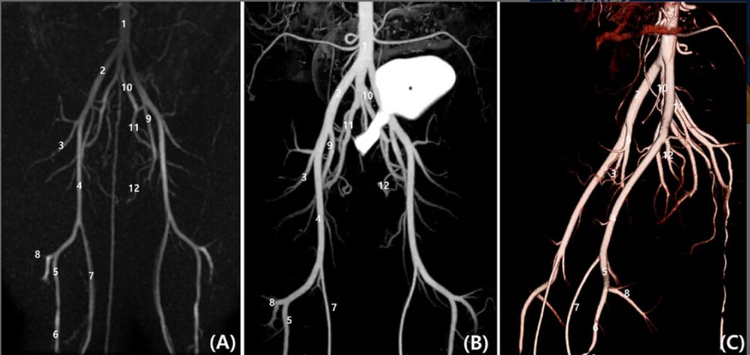
A, Dorsal planes Time-Resolved Imaging of Contrast Kinetics (TRICKS), (B) postprocessed subtraction CT angiography (CTA) maximum intensity projection image, and (C) sagittal plane of three-dimensional reconstructed CTA images (C). The subtraction reconstruction image was generated using a semi-automatic bone-subtraction method in a software program (Xelis, Infinitt). Both TRICKS and CTA demonstrate the following arterial branches: aortic bifurcation (1), external iliac (2), lateral circumflex femoral (3), femoral (4), popliteal (5), cranial tibial (6), saphenous (7), distal caudal femoral (8), deep femoral (9), internal iliac (10), gluteal (11), and internal pudendal arteries (12). The asterisk (*) indicates the urinary bladder with contrast medium.
How did we do? |
Disclaimer: The summary generated in this email was created by an AI large language model. Therefore errors may occur. Reading the article is the best way to understand the scholarly work. The figure presented here remains the property of the publisher or author and subject to the applicable copyright agreement. It is reproduced here as an educational work. If you have any questions or concerns about the work presented here, reply to this email.