- Veterinary View Box
- Posts
- What happens to the prostate long term post castration?
What happens to the prostate long term post castration?
Frontiers in Veterinary Science 2025
Stefano Spada, Daniela De Felice, Sebastian Arlt, Luiz Paulo Nogueira Aires, Gary C. W. England, Marco Russo
Background
Castration is widely used for population control and prevention of androgen-dependent diseases in male dogs, such as benign prostatic hyperplasia (BPH). However, while short-term prostatic involution has been documented within the first 90 days post-castration, little is known about long-term changes in prostate size and vascularization. This study aimed to assess the ultrasonographic and contrast-enhanced ultrasound (CEUS) changes in the prostate gland six years post-castration.
Methods
Study Design:
-10 adult neutered dogs were evaluated at two time points:
-T0 (initial assessment): at least 3 months post-castration.
-T1 (follow-up): six years later.
Ultrasound Evaluation:
-B-mode ultrasound: Prostate size, shape, margins, echogenicity, and echotexture were assessed.
-Prostate volume was calculated using Atalan’s formula.
Contrast-Enhanced Ultrasound (CEUS):
-The contrast agent SonoVue was administered intravenously.
Perfusion parameters measured:
-Peak perfusion intensity (PPI) (% contrast enhancement).
-Time to peak perfusion (TTP) (seconds).
Statistical Analysis:
-Wilcoxon signed-rank test compared prostate volume and perfusion between T0 and T1.
Results
B-mode Findings:
-The prostate remained hypoechoic and homogeneous, with minimal volume reduction (median 7.55 cm³ at T0 to 7.39 cm³ at T1, p = 0.005).
-No significant echogenicity or structural changes were observed over time.
CEUS Findings:
Prostate perfusion significantly declined over six years:
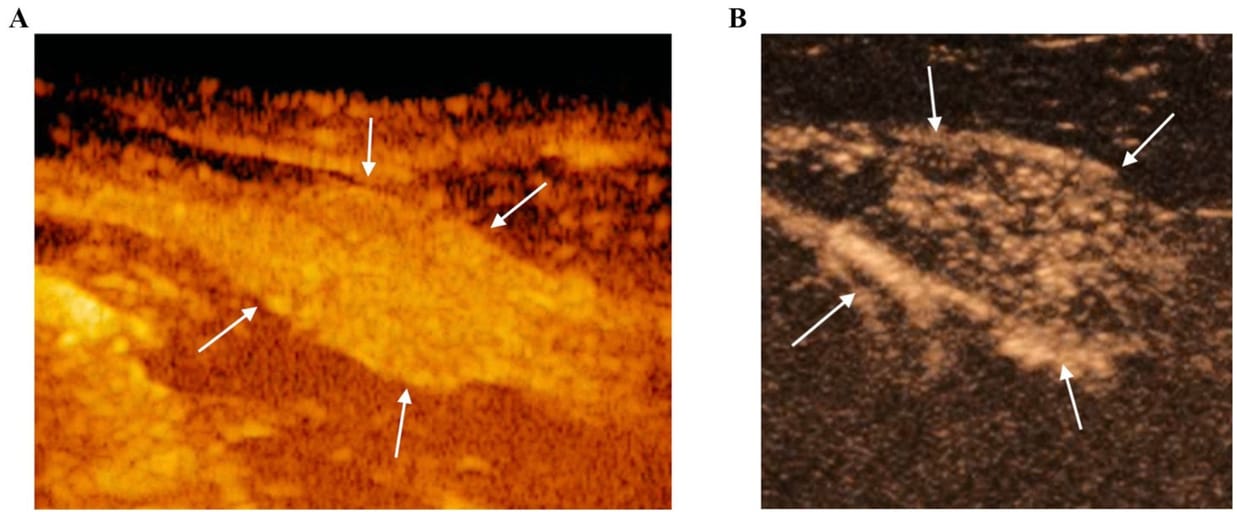
-PPI decreased from 54.9% at T0 to 29.6% at T1 (p = 0.005).
-TTP increased from 26.3 s to 47.0 s (p = 0.005).
-The reduction in vascularization was more pronounced in larger dogs (p = 0.028).
Limitations
Small sample size (n = 10), limiting generalizability.
No control group of intact male dogs for comparison.
Only two time points (T0 and T1), preventing detailed tracking of gradual involution.
Conclusions
Prostatic involution continues beyond the first 90 days post-castration, with significant vascular regression observed over six years. CEUS detected marked reductions in perfusion, which were not apparent on B-mode ultrasound. These findings highlight that castration-induced changes in prostate physiology are prolonged and may influence the risk of prostatic neoplasia. Further research is needed to correlate perfusion changes with potential neoplastic transformation in neutered dogs.

CEUS of the prostate gland (white arrows) at the peak enhancement performed at T0 (A) and T1 (B). At T1 the prostate gland resulted to be less homogeneous and characterized by a reduced contrast intensity when compared to T0 by using CEUS
How did we do? |
Disclaimer: The summary generated in this email was created by an AI large language model. Therefore errors may occur. Reading the article is the best way to understand the scholarly work. The figure presented here remains the property of the publisher or author and subject to the applicable copyright agreement. It is reproduced here as an educational work. If you have any questions or concerns about the work presented here, reply to this email.