- Veterinary View Box
- Posts
- What is the cause of linear cystic calculi?
What is the cause of linear cystic calculi?
Veterinary Radiology & Ultrasound, 2020.
Jennifer M. Hickey, Allyson C. Berent, Anthony J. Fischetti, Alexandre B. Le Roux.
Background
Cystic calculi in dogs can arise from various causes, including diet, infection, metabolic factors, and urinary tract foreign bodies. Suture-associated calculi have been documented as an uncommon complication of cystotomy, potentially occurring due to suture material acting as a nidus for mineralization. The objective of this study was to describe the radiographic features of suspected suture-associated cystic calculi in dogs with a history of prior cystotomy.
Methods
This retrospective case series reviewed medical records and radiographs of 644 dogs and cats that underwent cystolith removal over a seven-year period. Inclusion criteria required prior surgical stone removal, presence of calculi with a hollow center on gross analysis, and availability of radiographic images. Radiographs were analyzed by two veterinary radiologists and an internal medicine specialist.
Results
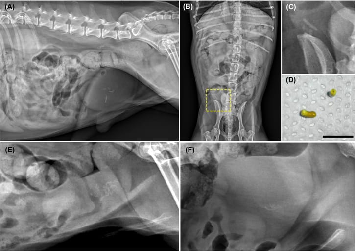
Seven cases were identified, involving six dogs (one dog presented twice). Affected dogs were all neutered males, small breed (3.2–10.7 kg), and middle-aged (mean 9.4 years). Radiographic findings showed multiple linear, mineral-dense calculi located centrally within the bladder. One dog exhibited both round and linear calculi. Gross examination confirmed a hollow core in all cases. Calculi were composed of calcium oxalate. Prior cystotomies had used monofilament absorbable sutures (polydioxanone or poliglecaprone 25), though two cases had unknown suture material. Recurrence time ranged from 3 months to 2 years post-surgery.
Limitations
The study was limited by its small sample size, retrospective nature, and lack of statistical analysis. Additionally, the specific role of different suture materials in calculus formation could not be fully assessed.
Conclusions
Suture-associated cystic calculi should be considered in dogs with a history of cystotomy, especially when radiographs reveal linear, centrally located, mineral-dense calculi. To minimize risk, surgeons should avoid placing suture material within the bladder lumen and consider alternative calculus removal techniques such as voiding urohydropropulsion or minimally invasive procedures.

A, Right lateral abdominal compression study (digital radiography, Canon CXDI-50 IP, kVp 90, mAs 4.0) in a 10-year-old MN Poodle Mix. This study increases conspicuity of multifocal, linear, mineral opacities within the urinary bladder. Pin point mineral foci are also present within one of the kidneys. B, ventrodorsal projection (digital radiography, Canon CXDI-50 IP, kVp 90, mAs 4.0) of the abdomen of the same patient. Multifocal, linear, mineral opacities are within the urinary bladder, located to the right of mid line. C, Cropped image of multifocal cystic calculi on the previous ventrodorsal projection. D, Gross image of suture-related cystic calculi associated with the prior percutaneous cystolithotomy site of the patient in A, B, and C. The cystic calculus is cylindrical in shape with a hollow core. The black bar indicates 0.5 cm in length. E, A right lateral abdominal cropped image (digital radiography, Canon CXDI-50 IP, kVp 90 mAs 4.0) in a 7-year-old MN Pomeranian with round, pin point as well as linear mineral opacities within the urinary bladder. F, A right lateral abdominal cropped image (digital radiography, Canon CXDI-50 IP, kVp 90, mAs 4.0) in an 8-year-old MN Maltese Mix with multifocal linear mineral opacities within the urinary bladder [Color figure can be viewed at wileyonlinelibrary.com]
How did we do? |
Disclaimer: The summary generated in this email was created by an AI large language model. Therefore errors may occur. Reading the article is the best way to understand the scholarly work. The figure presented here remains the property of the publisher or author and subject to the applicable copyright agreement. It is reproduced here as an educational work. If you have any questions or concerns about the work presented here, reply to this email.